Robust data now document that the more time an individual spends on social media, the more likely they will be bullied, and the vast majority of all social media users have seen severe cyberbullying. One heartbreaking example that made national news recently is of a teenage girl who was viciously attacked at school and subsequently committed suicide after videos of the incident turned up online.
One common reaction to such traumatic social stress is the development of anhedonia—a loss of interest in pleasurable activities—that can manifest within social domains and lead to severe social avoidance. Studies in humans have shown that social stress elicits feelings of social isolation and reduced responses to the rewarding aspects of social interactions, both at the behavioral and neural levels. (See related article on the neural circuit mechanisms of social isolation.)
To better understand the biological and psychological underpinnings of trauma-induced impairments in social behavior, researchers at The Friedman Brain Institute are using rodent models of social stress.
In one model, termed social defeat stress, a test mouse is exposed to a bigger, more aggressive mouse. After a brief physical interaction, the mice are separated by a screen but the test mouse continues to be subjected to aggressive behaviors by the bigger mouse. This process is repeated with a different aggressive mouse for several days. After such exposure, the test mouse exhibits profound social avoidance along with abnormalities in appetite and feeding behavior, body weight, sleep, and other behaviors reminiscent of human depression and anxiety disorders.
An important feature of the social avoidance induced by social defeat, which indicates that it is pathological, is that the test mice generalize it to littermates—mice with which they were raised. Another important feature is that these social deficits are essentially permanent and do not resolve even after the mice are rehoused with their former cage mates. Several teams at Mount Sinai have characterized regions of the brain that mediate the lasting deleterious consequences of social defeat stress along with the molecular changes that occur in those regions.
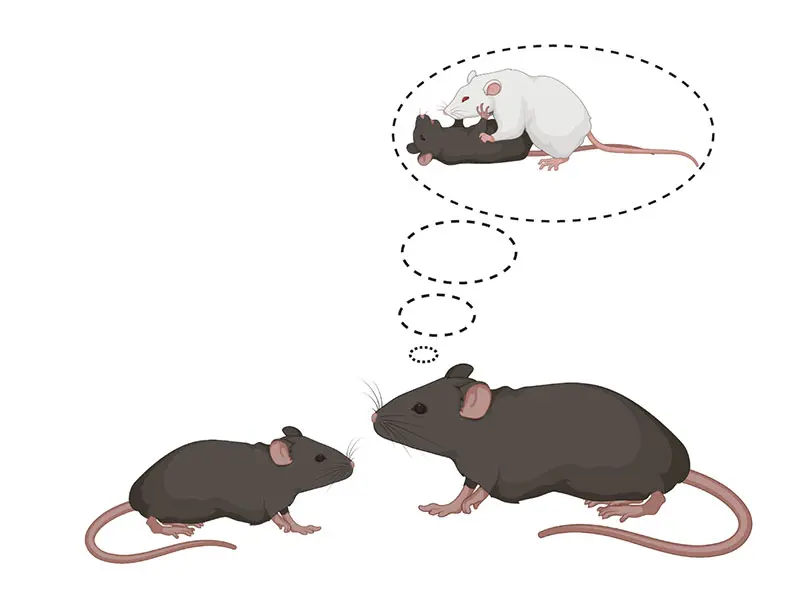
Figure 1: Past social trauma affects social behavior. The schematic shows that susceptible mice with a history of social trauma exhibit generalized social avoidance behavior, not only to traumatic social clues, but also to rewarding social clues (such as nonaggressive juveniles). Created with BioRender.com.
A leader of these investigations is Scott Russo, PhD, Mount Sinai Professor in Affective Neuroscience, Professor of Psychiatry, Director of the Center for Affective Neuroscience, and the Leon Levy Director of the Brain and Body Research Center at the Icahn School of Medicine at Mount Sinai. Recent work from the Russo Laboratory found that mice that underwent social defeat stress even avoid nonthreatening juvenile mice (see Figure 1), which is highly unusual, since interactions with juvenile mice, in contrast to aggressive mice, are especially rewarding and sought after.
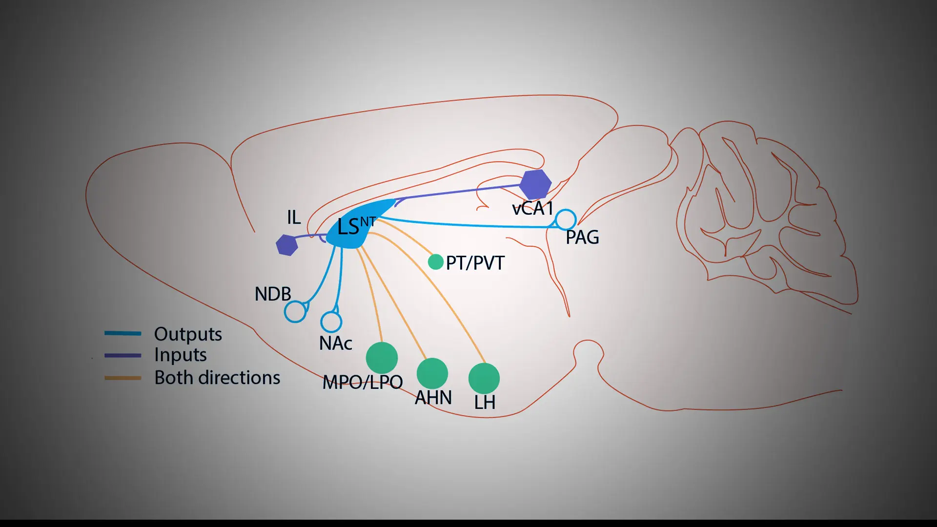
The researchers identified a region of the brain called the lateral septum, located within the subcortical forebrain that has long been known to respond to stress and threats in the environment, which is highly activated in these defeated mice when they interact either with aggressive mice or nonthreatening juvenile mice. These findings show that the repeated bullying experienced during social defeat “rewires the brain” in ways that make normally rewarding social targets seem stressful or threatening, thereby engaging the lateral septum circuitry to impair social reward (see Figure 2).
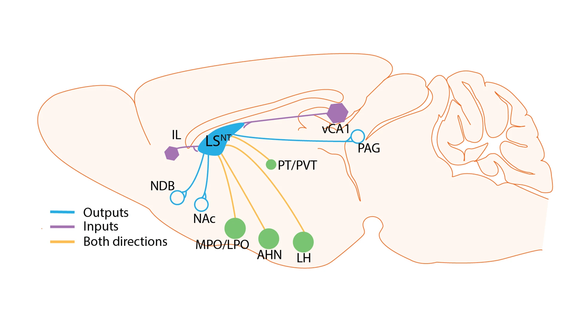
Figure 2: Input-output map of lateral septum (LS) neurons. The schematic focuses on a subset of LS neurons that express the neuropeptide, neurotensin (NT), and shows the prominent connections of these cells with other brain regions. Lateral septum neurotensin neurons project to the medial preoptic area/lateral preoptic area (MPO/LPO), paraventricular thalamus (PVT), paratenial thalamus (PT), anterior hypothalamic nucleus (AHN), lateral hypothalamus (LH), nucleus accumbens (NAc), nucleus of the diagonal band (NDB), and periaqueductal gray (PAG). Lateral septum neurotensin neurons also receive input from ventral CA1 of hippocampus, infralimbic cortex (IL), and MPO/LPO, AHN, and LH.
Interestingly, in studies of patients with depression and comorbid social anxiety disorder, social trauma can increase the representation of social threat. Children who have experienced bullying and other social traumas exhibit heightened sensitivity to perceived threats, erroneous classification of neutral emotional stimuli, and attention biases toward threat-related cues. Future studies in humans will confirm the ways in which the lateral septum circuitry mediates social threat perception and reward sensitivity in victims of trauma, as seen in the mouse model, and such work has the potential to inform new therapeutic strategies for disturbances in social behavior that characterize many psychiatric syndromes.
Featured

Scott Russo, PhD
Mount Sinai Professor in Affective Neuroscience; Professor of Psychiatry; Director, Center for Affective Neuroscience; and Leon Levy Director, Brain and Body Research Center