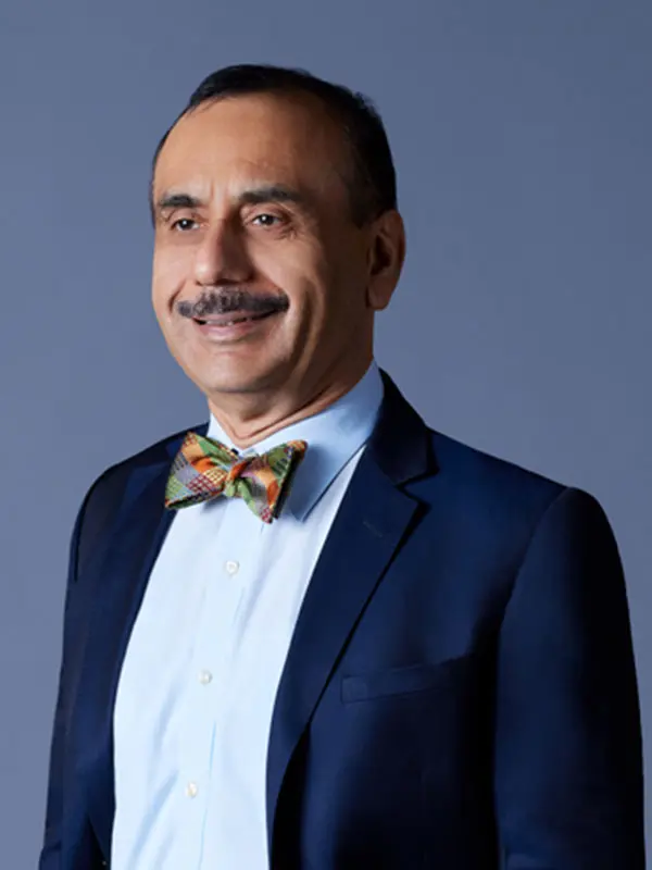
During a fall 2021 conference, Ash Tewari, MBBS, MCh, FRCS (Hon.), who has been a pioneer in robot-assisted radical prostatectomies (RARP) and related anatomical studies. reflected on these innovations and their benefits for patients who present with prostate cancer.
For more than a decade, Ash Tewari, MBBS, MCh, FRCS (Hon.), Professor and System Chair, Milton and Carroll Petrie Department of Urology, and Director of the Center of Excellence for Prostate Cancer at The Tisch Cancer Institute at the Icahn School of Medicine at Mount Sinai, has been a pioneer in robot-assisted radical prostatectomies (RARP) and related anatomical studies. Through this work, which includes more than 8,000 surgeries, he has introduced and refined several innovative nerve-sparing techniques and anatomical models that have struck a unique balance between optimal oncological outcomes and maintenance of continence and sexual function among patients.
Dr. Tewari’s focus on precision medicine, scientific innovation, and offering patients more options exemplifies Mount Sinai’s leadership in translating insights from research to clinical practice. During a fall 2021 conference, Dr. Tewari reflected on these innovations and their benefits for patients who present with prostate cancer.
An innovator in the fields of robot-assisted prostate cancer surgery and prostate cancer treatment, Dr. Tewari is a pioneer in the development of an athermal and traction-free technique during robotic prostatectomy to minimize the damage to the nerves responsible for erectile function. He developed the total reconstruction technique, which involves restoration of the support structures of the urinary mechanism following prostate removal, for faster continence recovery. In addition, he was the first to perform a catheter-less robotic prostatectomy where a urethral catheter is avoided to minimize pain and discomfort after surgery.
Drawing on his extensive anatomical background, Dr. Tewari was the first to describe the nerves responsible for erectile function as a neural hammock. He also described various grades of nerve-sparing that utilize information from MRIs and allow for incremental nerve-sparing—even in cases that would not have qualified for nerve-sparing without this graded approach. This increased understanding of the prostate anatomy aids in nerve-sparing prostate surgery techniques. Using a multiparametric magnetic resonance imaging (mpMRI)-based nomogram, Dr. Tewari can customize his approach to nerve-sparing based on a particular patient’s biology and pathology to achieve optimal continence, erectile function, and oncological outcomes.
He has described traction-free and athermal nerve-sparing techniques and has developed a novel hood technique for early urinary continence, which changed the approach surgeons used because it offers comparable results for urinary insentience and is easier than the traditional posterior approach. Working with genitourinary pathologists and scientists from the University of Hamburg, he helped advance the role of MRI-guided NeuroSAFE® and sphincter-safe approaches in expanding opportunities for nerve sparing.
As an active researcher and surgeon scientist, Dr. Tewari is one of only a few robotic surgeons to be awarded a National Institutes of Health R01 and Department of Defense (DOD) cancer grant. This funding is for the investigation of real-time tissue identification during prostate cancer surgery and immunotherapy.
Novel Nomogram for Predicting Seminal Vesicle Invasion and Extracapsular Extension
Traditionally, one of the key challenges in balancing oncological outcomes and the maintenance of continence and sexual function among patients undergoing RARP has been seminal vesicle invasion (SVI) and extracapsular extension (ECE). Dr. Tewari led the development of a side-specific predictive SVI and ECE model to guide presurgical decision-making related to nerve sparing based on four variables: prostate-specific antigen, highest ipsilateral biopsy Gleason grade, highest ipsilateral percentage core involvement, and seminal vesical invasion and ECE on multiparametric magnetic resonance imaging (mpMRI). A study involving data from 544 patients confirmed that the model is easy to apply in clinical practice, reproducible, and is better at predicting SVI and ECE than using mpMRI alone.
“These nomograms, which have been built by my group, enable us to assess the likelihood of an extra capsular extension that may not be visible on an MRI,” Dr. Tewari says. “We are constantly expanding, evaluating, and validating them to get a better sense of the grade of nerve sparing we can attempt on each side of the prostate. It is a very risk-stratified approach to achieving all three therapeutic goals.”
Hood Technique
Pioneered by Dr. Tewari, the hood technique uses an anterior approach to preserve the contents of the space of Retzius during RARP. Upon removal of the prostate, the preserved tissue has a “hood-like” appearance, comprised of the detrusor apron, arcus tendineus, puboprostatic ligament, anterior vessels, and some fibers of the detrusor muscle. The hood effectively encloses and protects the membranous urethra, external sphincter, and supportive structures.
A study led by Dr. Tewari found that, among 300 patients who underwent RARP with the hood technique, 62 percent achieved continence one week after removal of their catheter and 95 percent achieved continence at eight weeks.
“Our technique enables surgeons to preserve the anterior structures that are crucial to restoring early continence following radical prostatectomy with low positive surgical margin rates and enhanced ability to visualize anatomical landmarks,” Dr. Tewari says. “It also enables surgeons to avoid the residual cancer typically associated with achieving early continence, and thus spare patients the subsequent hormone or radiation treatments that compromise their quality of life.”
Neural Supply of Urethral Sphincter
One of Dr. Tewari’s major milestones in advancing the safety, efficacy, and reproducibility of nerve sparing techniques has been his efforts to understand and map out the neuronal networks that surround the prostate. In addition to conceptualizing the neural hammock, he observed that there are incremental zonal compartments that enabled him to develop different degrees of nerve sparing based on the degree of extracapsular extension.
Dr. Tewari also helped demonstrate that the external urethral sphincter is innervated by autonomic and somatic nerves. Some autonomic fibers run posterolaterally within the neurovascular bundles; others run from the inferior hypogastric plexus to the posteromedial aspect of the prostate apex, above and through the rectourethral muscle. Furthermore, the external urethral sphincter is innervated by somatic nerves arising from the pudendal nerve, joining the midline but remaining below the rectourethral. Thus, the rectourethral muscle and the neurovascular bundles are to be preserved, particularly during apical dissection.
The Next Frontier
Having pioneered many nerve-sparing techniques for RARP and contributed to a better understanding of related neuronal networks, Dr. Tewari is now focused on the potential to enhance preprocedure planning through advances in imaging technology.
For example, he is looking into converting T2-weighted imaging, diffusion-weighted imaging, and dynamic contrast enhancements into 3D models that could facilitate decision-making related to the gradation of nerve sparing that is achievable in each patient. He is exploring the possibility of applying high-resolution 7-Tesla functional magnetic resonance imaging to glean further anatomical insights. Additionally, he is analyzing the potential use of artificial intelligence to gather information to create models that could predict, for example, the risk of SVI and ECE among patients. He is also exploring the potential of using multiphoton microscopy for real-time visualization of nerves and extent of ECE among patients.
“It will not be too far in the future that we will be able to see a very high-resolution image of cancer and where the nerves are, and this level of imaging will take us to the next frontier in terms of the outcomes we can achieve for our patients,” Dr. Tewari says.
Featured

Ash Tewari, MBBS, MCh, FRCS (Hon.)
Professor and System Chair, Milton and Carroll Petrie Department of Urology, Director of the Center of Excellence for Prostate Cancer at The Tisch Cancer Institute at the Icahn School of Medicine at Mount Sinai, and Surgeon-in-Chief of the Tisch Cancer Hospital at The Mount Sinai Hospital