As science has begun to demonstrate, measuring retinal and choroidal blood flow to the eye can provide extraordinary clues not only to ocular diseases like glaucoma, but to systemic abnormalities that include diabetes and cardiovascular, sickle cell, and Alzheimer’s diseases. The hitch is that measuring localized blood perfusion has for years presented a real challenge, with no device providing the level of precision scientists and clinicians need.
That could soon be changing thanks to new technology known as Doppler holography, which New York Eye and Ear Infirmary of Mount Sinai (NYEE) is clinically developing in partnership with a handful of research centers around the world. Over the past year, NYEE has assembled a Doppler holography imaging system on-site and started using it to acquire data from patients that will be pivotal to seminal studies of the sophisticated technology now getting underway. Helping to propel the work is a $100,000 grant from the New York Eye and Ear Infirmary Research Foundation.
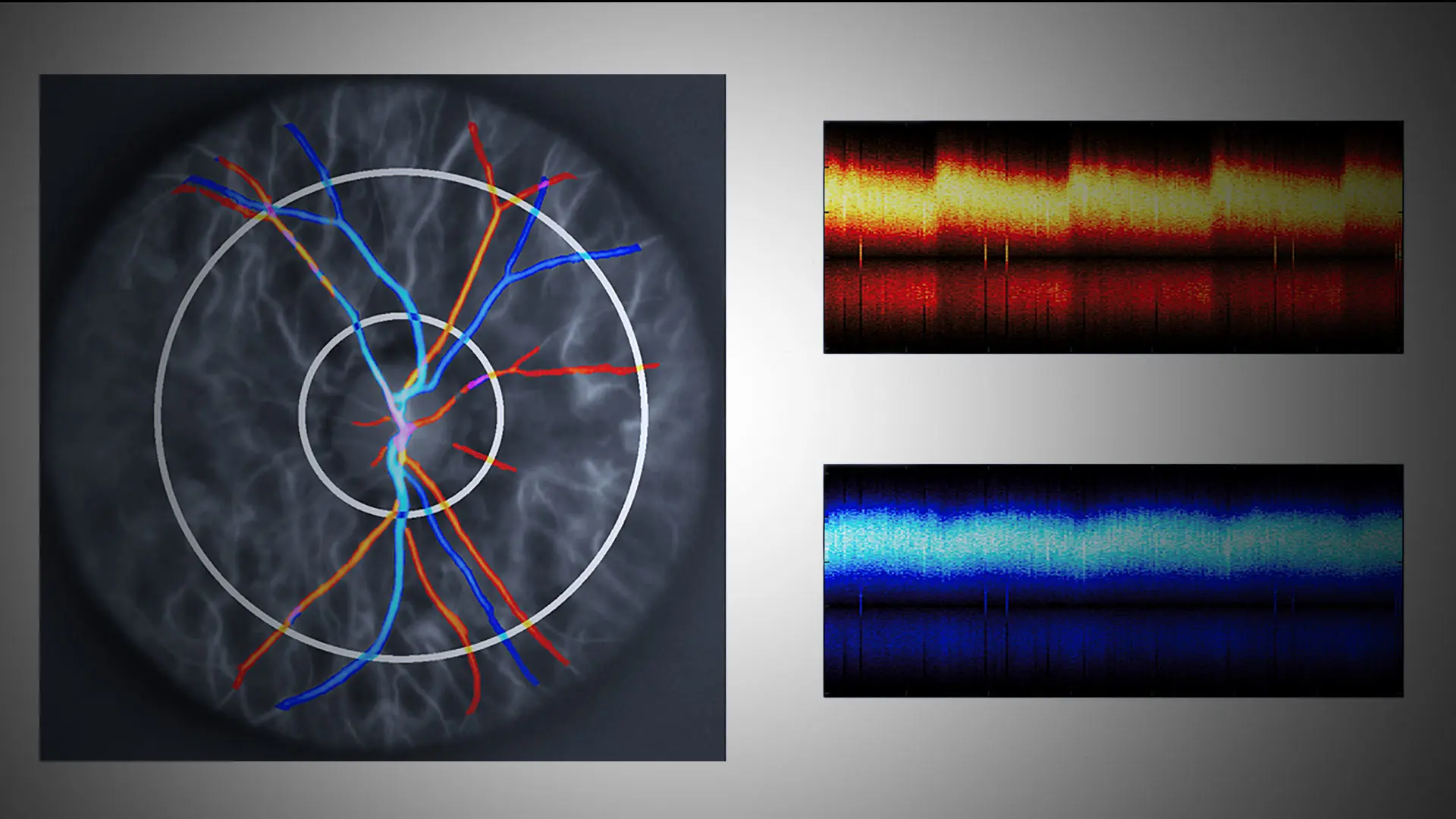
“We’ve been getting very exciting images from Doppler holography thanks to its ability to accurately measure total flow of blood into and out of the optic nerve,” says Richard B. Rosen, MD, FARVO, Vice Chair of Ophthalmic Research at NYEE, who is spearheading the hospital’s research in this field. “It will provide us with an important metric for diagnosing many diseases by offering clues about a variety of perfusion deficits to the eye that are characteristic of conditions like glaucoma and retinal vascular diseases. We are particularly interested in using the technology to detect early changes that occur in our patients with diabetes and sickle cell disease.”
Dr. Rosen, who is also the Belinda Bingham Pierce and Gerald G. Pierce, MD, Distinguished Professor of Ophthalmology at the Icahn School of Medicine at Mount Sinai, has looked at many different approaches to measuring blood flow in the retina over the years, including scanning laser ophthalmoscopy, stroboscopic fundus angiography, and most recently OCT Doppler imaging. The latest device being tested is a noninvasive holographic imaging technique fixed to a chin rest, like a slit lamp. However, unlike handheld Doppler ultrasound, it is more repeatable and less operator-dependent, producing whole-volume images of blood flow with the ability to focus on specific vessels within the retina. The system is powered by high-performance software that is consistently updated by the creator of Doppler holography—research investigator Michael Atlan, PhD, at the Institut Langevin Ondes et Images in Paris.
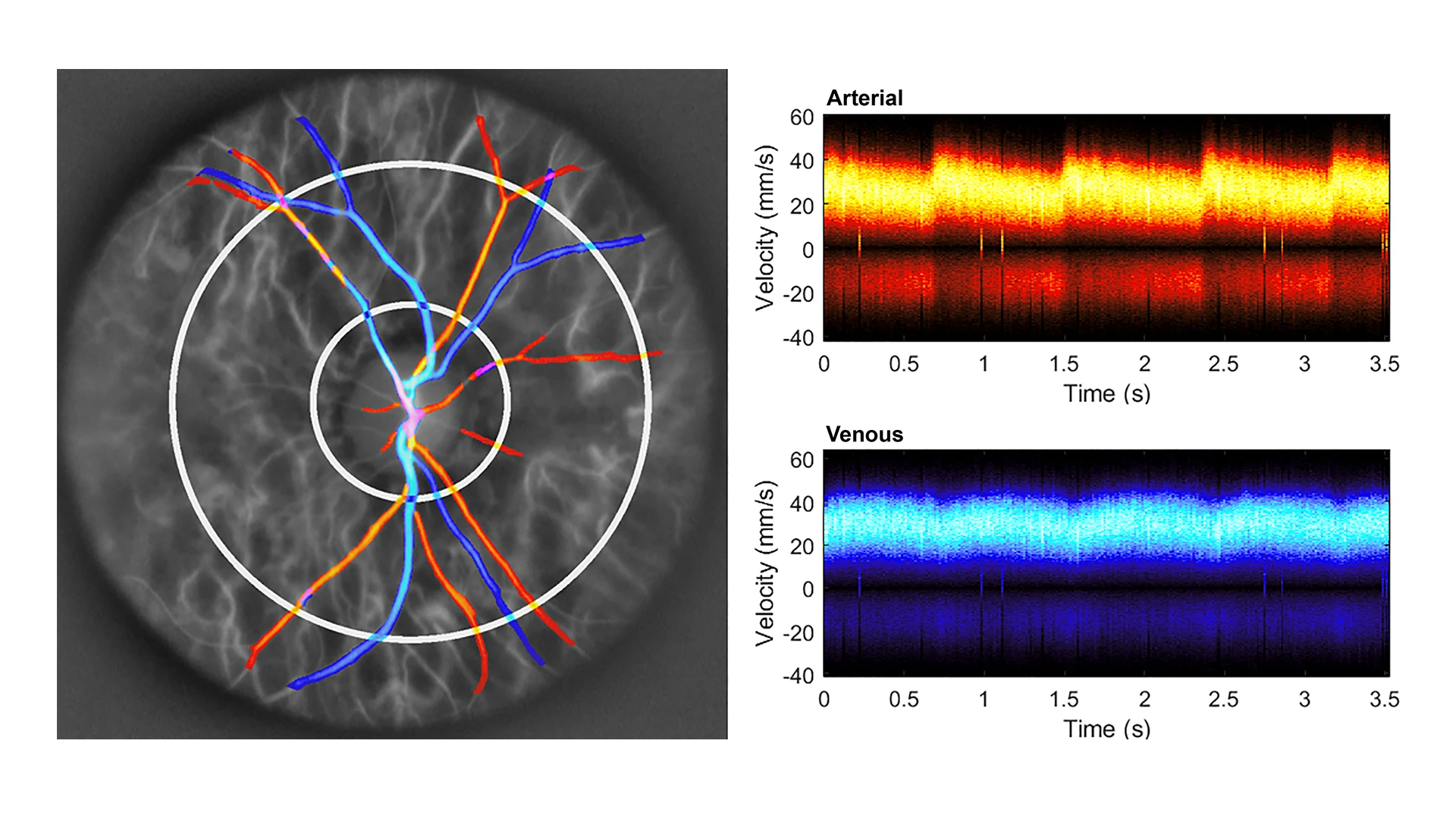
Healthy control, with normal intraocular pressure
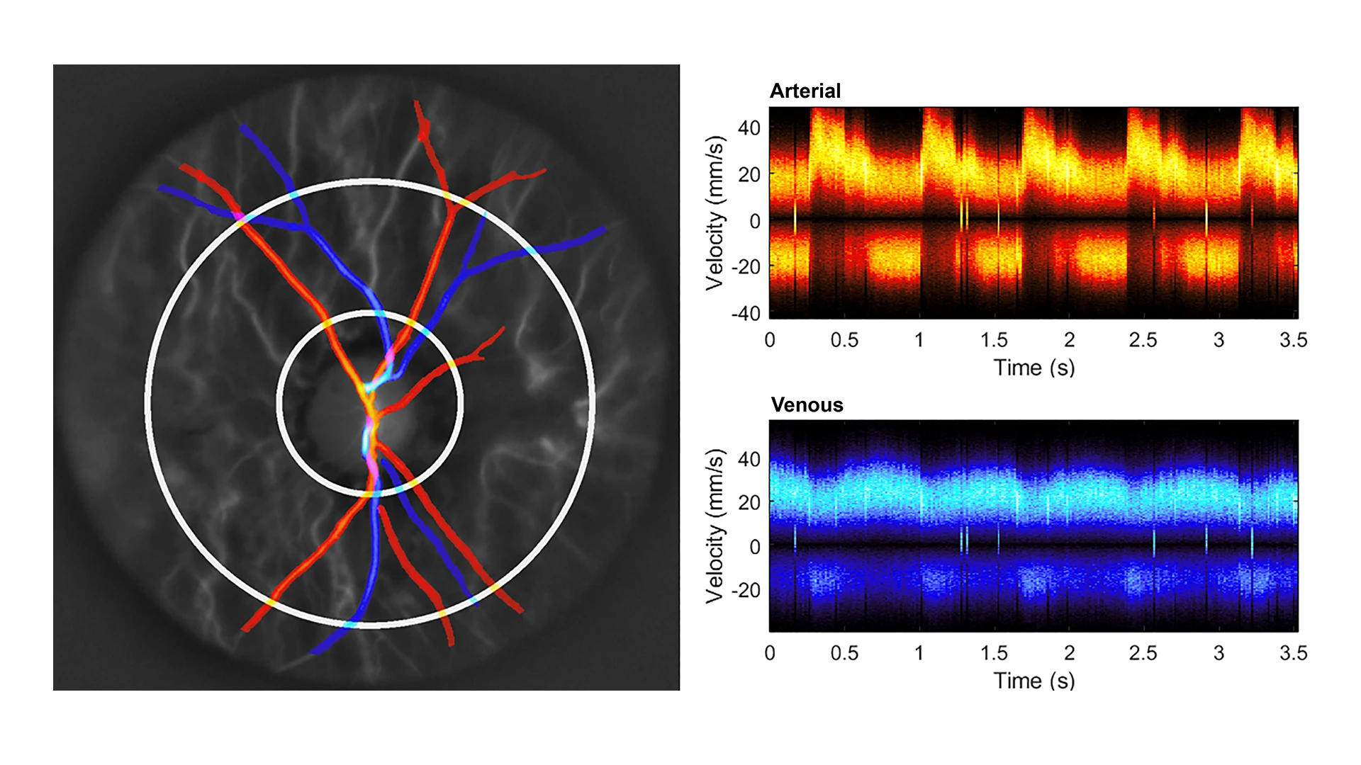
Same healthy control, exhibiting digitally elevated intraocular pressure
NYEE is now part of a global collaboration with Dr. Atlan and academic research centers in Europe, as well as a second U.S. site at the University of Pittsburgh, to clinically test the Doppler microscopy system. “We will all benefit from sharing our findings among the research partners,” acknowledges Dr. Rosen. “Our role now is to determine how we can best use Doppler holography to enhance patient care. To that end, we have been collecting normative data from healthy subjects in the lab and are now expanding our studies to patients with clinical problems.”
In addition to Dr. Rosen, Alon Harris, MS, PhD, FARVO, Professor of Ophthalmology, and Artificial Intelligence and Human Health, and Co-Director of the Barry Family Center for Ophthalmic Artificial Intelligence and Human Health at the Icahn School of Medicine, will figure prominently in the unfolding investigation into Doppler holography. The project is a perfect fit for Dr. Harris, who has focused his 25-year research career on the relationship between glaucoma prevalence and progression and vascular abnormalities. Dr. Harris is one of the pioneers worldwide to apply an ultrasound device called color Doppler imaging to measure blood flow dynamics in the human eye.
“Doppler holography could be useful in detecting many vascular-related diseases, and help identify their potential risk to patients,” says Dr. Harris. “It has many potential applications within the expanding field of oculomics, where we’re using powerful imaging techniques along with machine learning to identify the early presence of systemic pathologies including cardiovascular disease, hypertension, and diabetes.”
Dr. Rosen is equally bullish on the vast potential of Doppler holography. “We believe it could become an important tool for ophthalmologists, cardiologists, and other physicians in a couple of years,” he predicts. “By combining advanced optical imaging and computing, it will allow us to detect a wide range of diseases earlier in order to improve long-term outcomes.”