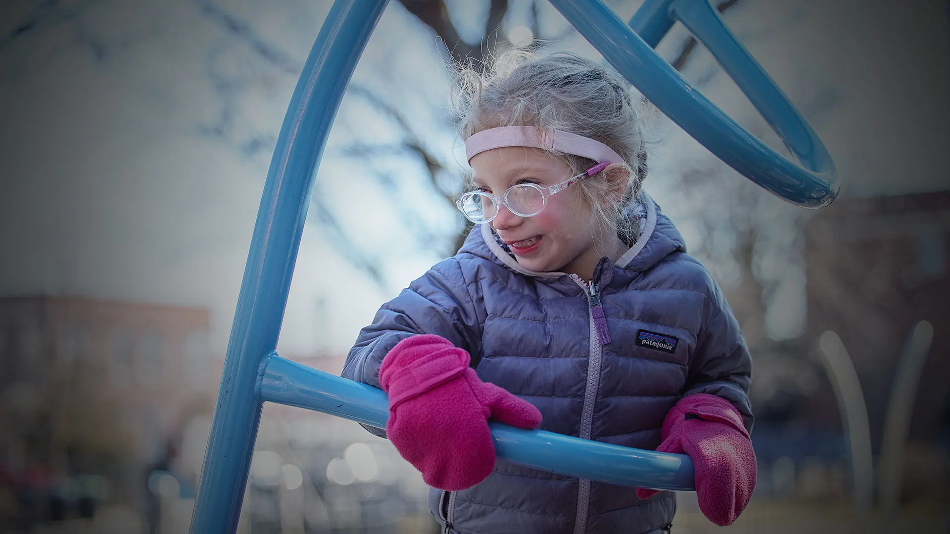
By three years of age, Avery Murynec had made up her mind to become an ophthalmologist. She certainly had no lack of role models.
The toddler, born with malformations of the right side of her face and eyelid, and her parents were no strangers to doctors’ offices and operating rooms. Many of those were at New York Eye and Ear Infirmary of Mount Sinai (NYEE), whose multidisciplinary specialists have addressed, nearly since Avery’s birth, such recurring issues as dermolipoma, eyelid coloboma, amblyopia, ectropion, astigmatism, and repeated ear infections.
At no point was the breadth and depth of that clinical brilliance on fuller display than in an eight-hour operation in September 2023, which brought together an oculoplastic surgeon, a cornea specialist, a pediatric ophthalmologist, and an otolaryngologist for a marathon day of surgery.
“It was amazing how they were able to coordinate all these specialists in one room to get such a complex job done,” remarks Avery’s father, Mark. “It took a tremendous load off our minds.”
Timing was of utmost concern for the team. “Because good vision is still forming in a child that young, any interruption can freeze the process in a less-than-perfect state,” says Tamiesha Frempong, MD, MPH, Assistant Professor of Ophthalmology at the Icahn School of Medicine at Mount Sinai, who has played a critical role in managing Avery’s ongoing care as her pediatric ophthalmologist. “For that reason, we knew we had to address her problems quickly.”
Indeed, the problems that confronted team members as they entered the surgical suite at The Mount Sinai Hospital last fall were manifold. Avery’s lower eyelid was turned out and essentially stuck to her cornea, making it difficult to close the eye. A large dermolipoma mass was present with scar tissue that had regrown on the surface of the cornea. Astigmatism aggravated by recurring scar tissue was compromising visual development of the affected right eye and, if not properly treated and managed, could lead to irreversible blindness in that eye.
“Aware of the high stakes involved, we made the collective decision to perform the multi-part, same-day surgery, including major eyelid reconstruction,” explains Valerie I. Elmalem, MD, Associate Professor of Ophthalmology at Icahn Mount Sinai and the oculoplastic surgeon on the case.
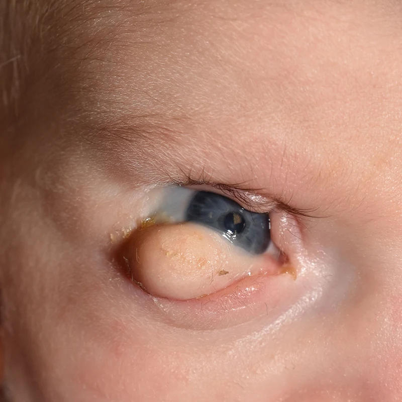
Appearance as an infant, age six months, prior to the first surgery on her eye; large inferior fornix lesion removed initially measured 2.2 cm x 1.5 cm x 0.4 cm
After first removal of the dermolipoma with no lesion on the cornea
Adds Neha Shaik, MD, a cornea specialist who, like each member of the team, remained in the OR and assisted others throughout the entire procedure: “Most hospitals would have had trouble handling this type of long and complex case, but everybody was on board and we came up with a common plan to enable this very young patient to hopefully see better for the rest of her life. And for me, it was fascinating to see the different specialties interacting so seamlessly.”
Dr. Shaik, an Assistant Professor of Ophthalmology at Icahn Mount Sinai, was familiar with Avery’s pathology. She had been part of the surgical team that operated on four-month-old Avery to help close a coloboma of the upper eyelid, as well as to remove the dermolipoma attached to the cornea and lower fornix of the inside of the eyelid.
Not unexpectedly, that scar tissue soon returned, fueling the patient’s astigmatism—and bringing Dr. Shaik back to the OR in September to again excise a benign mass on the inferior cornea. That fibrous tissue was now huge compared to the size of the eye itself, was deep-seated, and had grown multiple connections to not just the cornea but the skin and inferior fornix. Given its complexity, Dr. Shaik decided to perform a conjunctiva autograft once the dermoid was removed. Taking a small piece of tissue from the upper conjunctiva under the eyelid, Dr. Shaik transplanted it to the bottom part of the sclera, securing it in place with fibrin glue, followed by placement of a large amniotic membrane graft to promote healing and prevent recurrence of the inferior corneal mass.
The spotlight then turned to Dr. Elmalem’s reconstructive surgery to address the eyelid malformations, lower fornix, and orbital portions of the dermolipoma mass, and to try to prevent recurrence of the eyelid scarring to the cornea. To minimize damage to the eye muscle, Drs. Elmalem and Frempong worked together to remove the large inferior orbital portion of the dermolipoma that was attached to the inferior rectus muscle.
Also integral to this procedure was harvesting a mucous membrane graft from the inside of the patient’s mouth with the assistance of Aldo Londino III, MD, Assistant Professor of Pediatric Otolaryngology at Icahn Mount Sinai, allowing Dr. Elmalem to transplant it to the inside of the eyelid to basically recreate the conjunctival fornix. (Dr. Londino also played a key role in the day’s surgery by implanting ear tubes to guard against the patient’s frequent inner ear infections.) Dr. Elmalem applied amniotic membrane to the remaining regions of exposed sclera and affixed symblepharon rings under the upper and lower eyelids to keep the conjunctiva and eyelid from fusing with scar tissue. The ectropion was also repaired to allow better closure of the eyelids.
A postsurgical assessment underscores the importance of Avery’s omnibus surgery. “She still has astigmatism in the right eye, but it’s much better than it was before the dermoid was removed, and will continue to improve,” advises Dr. Frempong. “She’s also able to close her eye now, and vision in her right eye (now 20/70) is much better than before, when measuring acuity wasn’t even possible.”
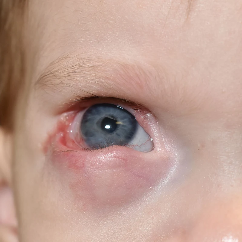
Regrowth of the dermolipoma/scar tissue onto the cornea with ectropion, age 2.5 years
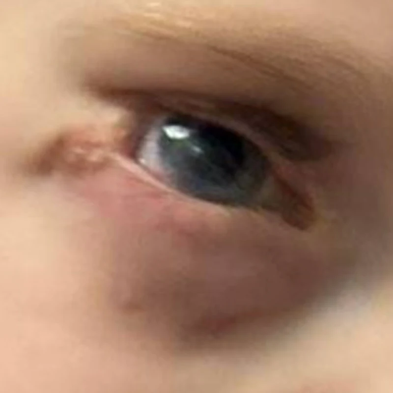
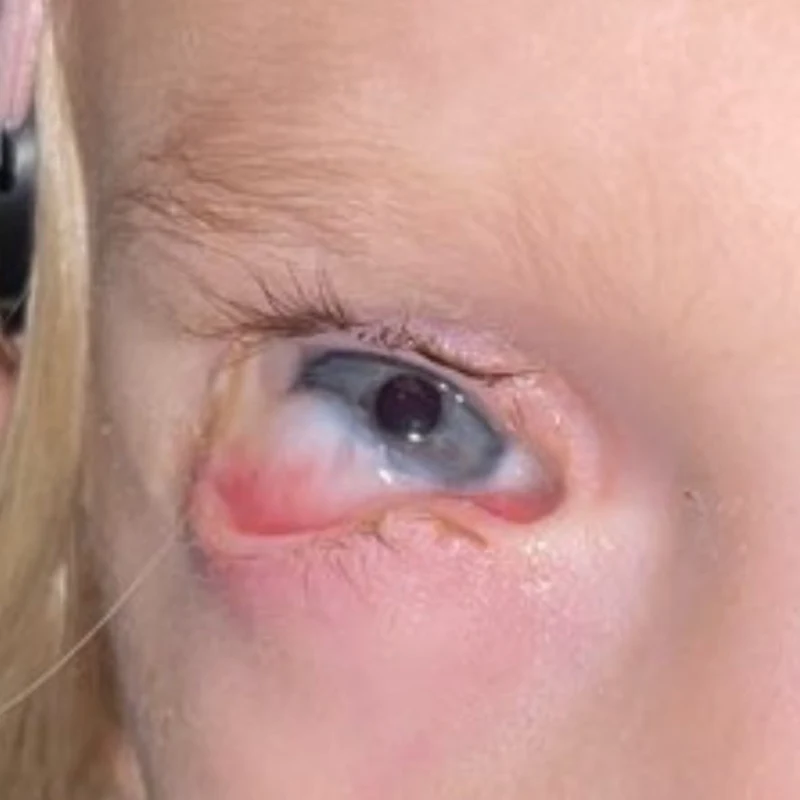
Four months after the second surgery, age 3 years 10 months. The cornea is clear and the lower eyelid is in good position.
Dr. Frempong’s intensive hands-on management includes working closely with the parents to ensure full-time use and the proper strength of corrective glasses, and the application of atropine drops and patching to “penalize” the good eye (which remains at 20/20) while promoting visual growth of the far weaker eye.
“The doctors have tempered our expectations of Avery’s left and right eyes ever looking or performing the same,” acknowledges Lauren, the patient’s mother. “But we’re hopeful that, as she grows and technology and surgical techniques advance, she will benefit from future opportunities. We feel extremely fortunate that NYEE has been part of our team at every step of the way, and that they’ll continue to be with us in the future.”
Featured
Valerie I. Elmalem, MD
Associate Professor of Ophthalmology
Tamiesha Frempong, MD, MPH
Assistant Professor of Ophthalmology
Neha Shaik, MD
Assistant Professor of Ophthalmology
Aldo Londino III, MD
Assistant Professor of Pediatric Otolaryngology