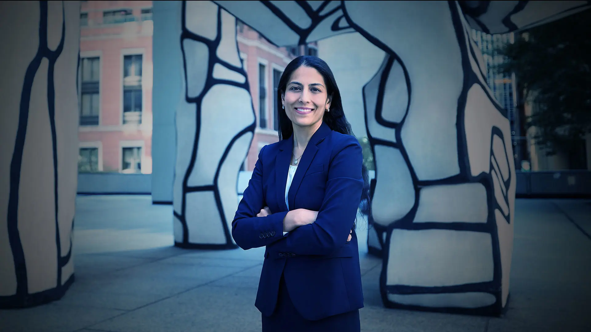
If Meenakashi Gupta, MD, Assistant Professor of Ophthalmology at the Icahn School of Medicine at Mount Sinai, has her way, the retina will become as critical a vital sign in annual physical exams of patients as temperature, blood pressure, pulse, and respirations.
And for good reason. A teleretinal screening program launched by New York Eye and Ear Infirmary of Mount Sinai (NYEE) in 2016 under her co-direction, now in place at seven primary care sites across the Mount Sinai Health System, has uncovered growing numbers of previously undiagnosed cases of diabetic retinopathy and other pathologies—particularly glaucoma and age-related macular degeneration—in patients undergoing their physicals.
According to a review of the model program from 2018 to 2022, nearly 15 percent of the 1,313 images taken in six participating doctors’ offices showed various stages of diabetic retinopathy. Moreover, 12.3 percent of those images also showed clinically significant macular edema, 9.8 percent showed glaucoma, and 8.1 percent revealed maculopathy.
“We are expanding teleretinal imaging throughout the Health System based on its success to date,” says Dr. Gupta, a vitreoretinal surgeon and Co-Director of Teleretinal Imaging. “When our imaging detects diabetic retinopathy, macular degeneration, or an optic nerve concerning for glaucoma, we notify the patient’s primary care doctor with recommendations for follow-up with an ophthalmologist.”
As of August 1, 2023, the teleretinal screening program, which provides for fundus cameras operated by trained medical personnel at each participating site, had evaluated more than 3,500 patients. The nonmydriatic images taken of the back of the eye are then securely transmitted to Dr. Gupta for later reading. While the program was originally targeted at diabetic patients at high risk for diabetic retinopathy, its lens is now widening to include other ocular disorders.
“As the program progresses,” reports Dr. Gupta, “we are finding there is much more pathology that we are able to capture, and hopefully at a stage where early intervention can prevent the patient’s condition from worsening.”
“As the program progresses, we are finding there is much more pathology that we are able to capture.”
- Meenakashi Gupta, MD
Beyond the growing caseload, what Dr. Gupta finds encouraging is the positive reception the program has received from the primary care community. “Providers are coming to us and requesting the program for their offices,” she notes. “An additional 30 sites could be onboarded in the near future across the metropolitan area.”
Another exciting prospect on the horizon for teleretinal imaging is artificial intelligence (AI). This unfolding technology has the potential to transform the program by using its algorithmic power to interpret fundus photographs at the point of care, providing immediate feedback to both providers and patients. The huge advantage is that if an abnormality is detected, an appointment with an ophthalmic specialist can be arranged before the patient leaves the office and is potentially lost to the system.
At present, AI only has Food and Drug Administration approval for use in diagnosing diabetic retinopathy. Technology needs to catch up to what the trained human eye can do. The ideal scenario for Dr. Gupta would be development of an algorithm with diagnostic ability to capture multiple pathologies.
“AI will definitely have a role in this program,” she advises. “We are now setting up the framework for that to happen, but we do not want to rush into it. We want to make sure we are using AI in the most responsible way for our patients.”
