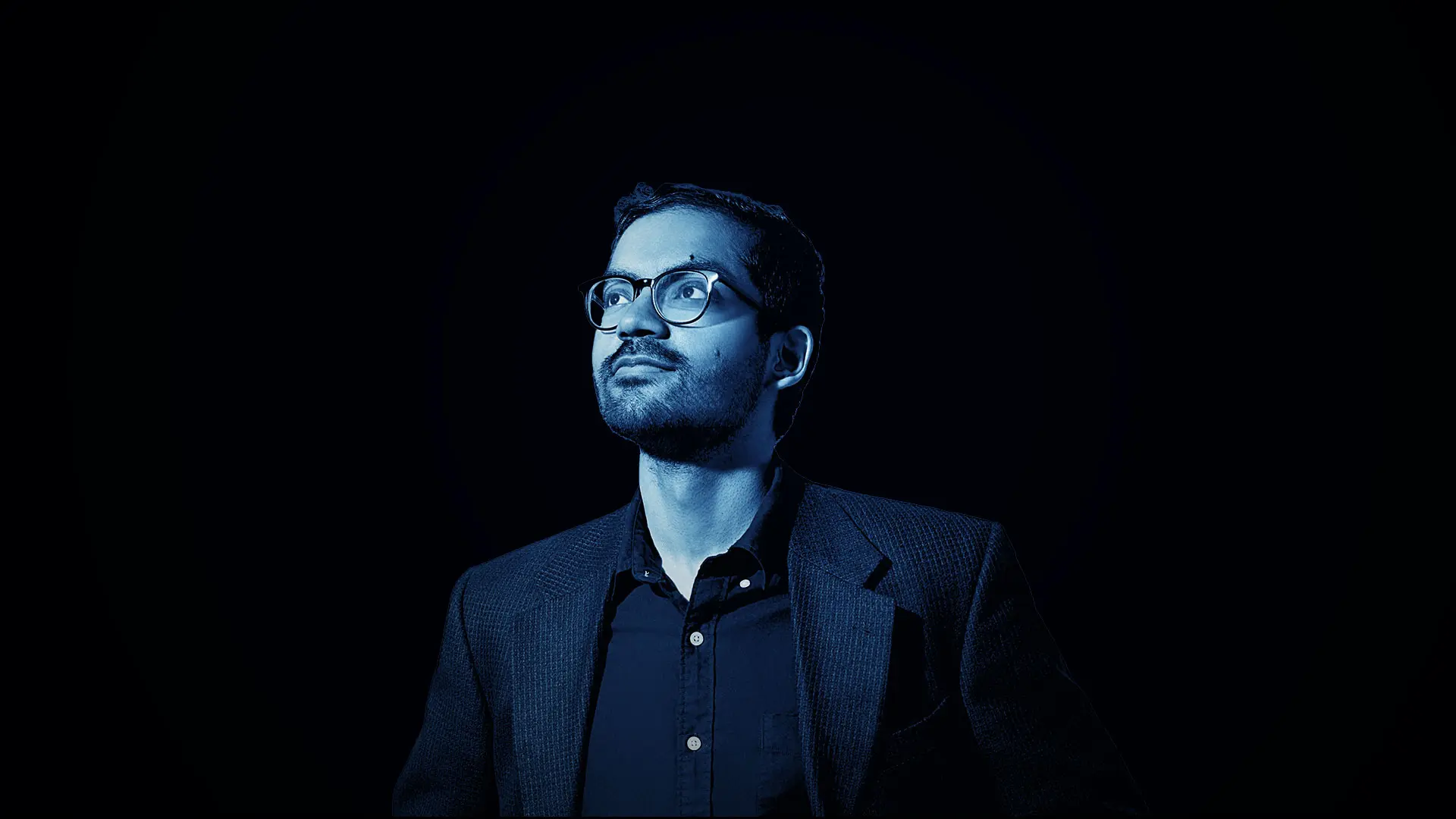
Treating eye injuries day after day at New York Eye and Ear Infirmary of Mount Sinai’s (NYEE) busy walk-in eye clinic in Manhattan reinforced for Shravan Savant, MD, the role of triage in ensuring patients the best outcomes. Just how important a role was driven home again to him by a 56-year-old man who entered the clinic one morning in March 2021 after a 10mm shard of metal from a machine shop penetrated his left eye, leaving him with intense pain and a traumatic wound in need of emergent care.
“With a complex case like this, the most immediate concerns are stabilizing the eye and triaging the patient in the most accurate and efficient way,” says Dr. Savant, who was a second-year resident when he first met the patient, Jan Gilewski. “My initial examination showed that the metal had pierced the patient’s cornea and reached back to the lens, so I immediately made plans to send him to Mount Sinai Beth Israel for a CT scan to check for other metal fragments in the posterior segment. At the same time, I developed a plan for surgery as soon as he returned.”
Given the nature of the wound, the No. 1 concern for Dr. Savant was infection, which could dramatically affect the outcome. “Apart from infection risk, a piece of metal in the retina is toxic, and can destroy vision very quickly,” he explains.
Of more immediate concern for Mr. Gilewski, a machine welder for New York City Transit, was the mounting pain. “I waited a bit, thinking it might go away, but when my vision became cloudy and the pain didn’t let up, I told my supervisor and we drove across the bridge to the clinic at New York Eye and Ear,” he recalls.
He was met by Dr. Savant, who soon began administering abundant amounts of antibiotic eyedrops, intravenous antibiotics, and tetanus vaccine to control the threat of infection. Meanwhile, the results of the CT, which arrived within an hour, showed the back of the patient’s eye to be free of any metal. With that welcome news, Dr. Savant and the voluntary attending, Luna T. Xu, MD, Assistant Clinical Professor of Ophthalmology, NYEE, began surgery late that afternoon. “The prognosis is always guarded when you have a large foreign body removal like this one,” explains Dr. Savant. “Even after the object is removed and the wound is closed, the patient faces the prospect of astigmatism, and vision out of the eye may never be as good as it originally was.”
When first attempts to push the object out by enlarging the wound opening proved unproductive, the surgeons used retinal forceps to snake the metal out from the front. That tactic allowed them to better assess the damage, and they learned that the force of the shard entering his eye had shattered its natural lens, triggering a traumatic cataract that precluded traditional lens removal. The capsule of the lens had split into the dreaded “Argentinian flag” configuration, preventing the normal techniques for capsulotomy. Instead, the surgeons used a technique known as a can opener capsulotomy in which they etch a circular series of tiny nicks in the anterior capsule surrounding the lens using a cutting instrument known as a cystotome, creating an opening to remove the cataract.
A case “shows what’s possible when you get the entire team involved, from radiology to our retina specialists to our on-call trauma attendings. We did everything by the book, and it resulted in an excellent outcome.”
- Shravan Savant, MD
With the cataract removed, the next challenge confronting Dr. Savant and Dr. Xu was selection and placement of a new intraocular lens. Once again this called for extraordinary measures since the foreign body protruding into the eye had damaged the capsular bag, the normal support structure for the lens. Instead, the team implanted a three-piece acrylic lens in the sulcus in front of the bag. That still left another critical step: repairing the gaping hole in the cornea where the metal had entered by suturing it closed.
The patient’s vision out of the injured eye improved to 20/50 within the first few days, and to nearly 20/20 within 10 days, with no signs of infection. “He’s actually seeing better out of his left eye than the other, which we never expected,” reports Dr. Savant. “It shows what’s possible when you get the entire team involved, from radiology to our retina specialists to our on-call trauma attendings. We did everything by the book, and it resulted in an excellent outcome.”
The patient would be the first to agree. “I knew NYEE was the best eye clinic in the city, and Dr. Savant confirmed that for me,” says Mr. Gilewski. “He dropped everything he was doing to focus on my case from the moment I walked in. He told me he would do everything he could to save my eye, and he certainly kept his word.”
