Among the debilitating effects of sickle cell disease, which often strikes in the prime of life, is sickle cell retinopathy, a condition that may go undetected until it has caused permanent damage to the eye. That picture has turned considerably brighter, however, with the development by researchers at New York Eye and Ear Infirmary of Mount Sinai (NYEE) of an innovative new technique for sequential imaging of retinal blood flow. In sickle cell patients, this technique can reveal how the disease is progressing, as well as the effectiveness of treatment regimens such as hydroxyurea. Their work was reported in a study published online in the May 2021 issue of Biomedical Optics Express.
“For the first time we’ve shown how OCT (optical coherence tomography) angiography can be used to evaluate the immediate status of sickle cell disease using dynamic retinal imaging that depicts microscopic changes in blood flow in the smallest blood vessels,” says Toco Chui, PhD, Director of the David E. Marrus Adaptive Optics Imaging Laboratory at NYEE, and senior author of the study. “This approach allows us to noninvasively monitor the retinal microcirculation over time and assess a sickle cell patient’s condition before or after initiation of therapy.”
Sickle cell disease is an inherited red blood cell disorder that afflicts about 10 percent of African Americans. A mutation in the hemoglobin gene causes the protein to fold abnormally, resulting in red blood cells that distort to a “sickled” shape when stressed. These misshapen cells clump and jam the blood flow in capillaries, which become inflamed and occluded. Progressive regions of blocked circulation can lead to loss of vision in the retina, which may ultimately result in blindness.
Currently, ophthalmologists rely on static images from single scans of the retina, a practice that fails to capture the dynamic nature of the disease process. OCT angiography provides access to the most delicate capillaries and the ability to measure and map microscopic vaso-occlusive events across short- and long-term intervals. In their analysis of 27 patients, NYEE researchers imaged each subject
10 times in a row over a 10-minute span. An hour later, they repeated the procedure to determine which blood vessels were repeatedly opening and closing. From these studies they were able to characterize the level of disease burden and the current activity of the sickle cell disease for the particular patient.
“We learned from our study that patients with no active sickle cell retinopathy show minimal intermittent fluctuation in capillary blood flow,” explains co-author Richard B. Rosen, MD, Chief of Retina Service, Distinguished Professor of Ophthalmology at the Icahn School of Medicine at Mount Sinai, and Vice Chair and Director of Ophthalmology Research at NYEE. “In patients with more frequent temporary capillary blockage there is probably a higher risk of permanent closure, which can lead to significant vision loss.”
This novel use of OCT angiography, a relatively new technology that NYEE has been in the vanguard of developing, could be an important tool in the armamentarium of clinicians who take care of patients with sickle cell disease. “It’s often impossible for these physicians to gauge the impending danger of the sickling condition until the patient experiences vision loss,” observes Dr. Chui. “That’s why OCT angiography could be a game changer, leading to earlier diagnosis of retinal issues and potentially avoiding irreversible blindness and vascular complications in other parts of the body.”
Researchers speculate that the technology could have implications that stretch well beyond sickle cell disease. “Microcirculation in the capillaries of the retina provides a unique window on what is happening in other parts of the body, like the brain and major organs,” says Dr. Rosen. “For that reason, OCT angiography could provide a strong platform for detecting a variety of circulation threats in a non-invasive way.”
[see below]
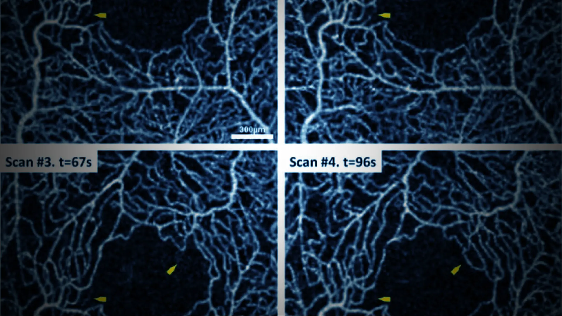
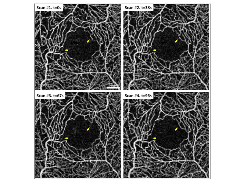
OCT Angiography scan sequence demonstrating intermittent capillary filling (frames 4 of 10).
This is a sickle cell HbSS disease patient on hydroxyurea treatment. Yellow arrows point to two capillary
segments bordering the foveal
avascular zone of the macula which demonstrate intermittent capillary
perfusion during the 10 scan sequence. The time of image acquisition is seen in the upper left corner of
each OCT-A scan.
[see below]
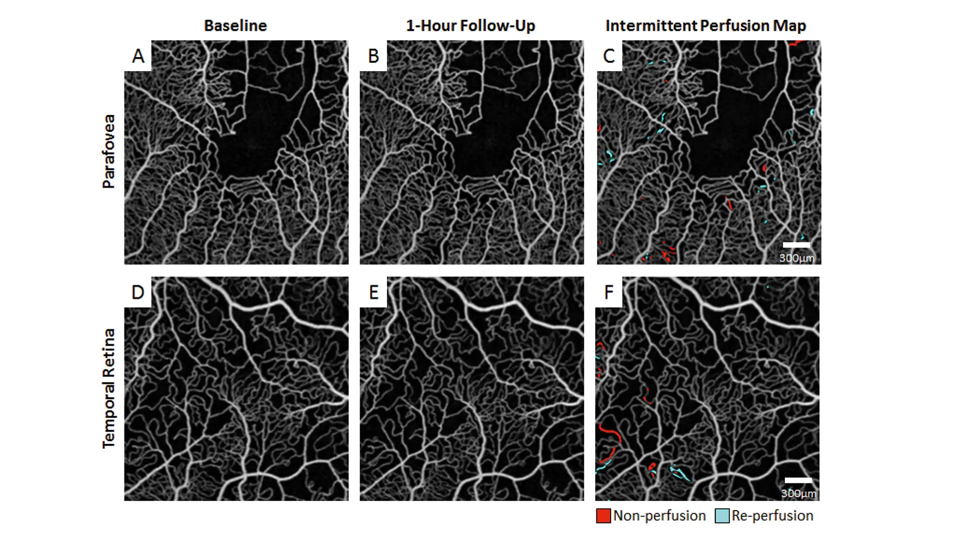
Labeling of non-perfused and reperfused capillaries. Averaged OCT angiography images of a sickle cell HbSC disease patient with non-perfused segments around the fovea (top row) and enlarged inter-capillary spaces in the temporal retina (bottom row) for the baseline (A & D) and 1-hour follow-up (B & E) imaging sessions. Intermittently filled capillary segments are labeled as non-perfusion (red) when they are filled at baseline but not at 1-hour follow-up (C & F). Non-filled segments at baseline which reappear at 1-hour follow-up are considered as reperfusion (cyan) (C & F).