The study findings, published online on September 3, 2025, in Neurosurgery, establish the safety of an approach that allows researchers to collect valuable living human brain tissue during planned neurosurgical procedures, addressing a critical barrier in neuroscience research while maintaining patient safety.
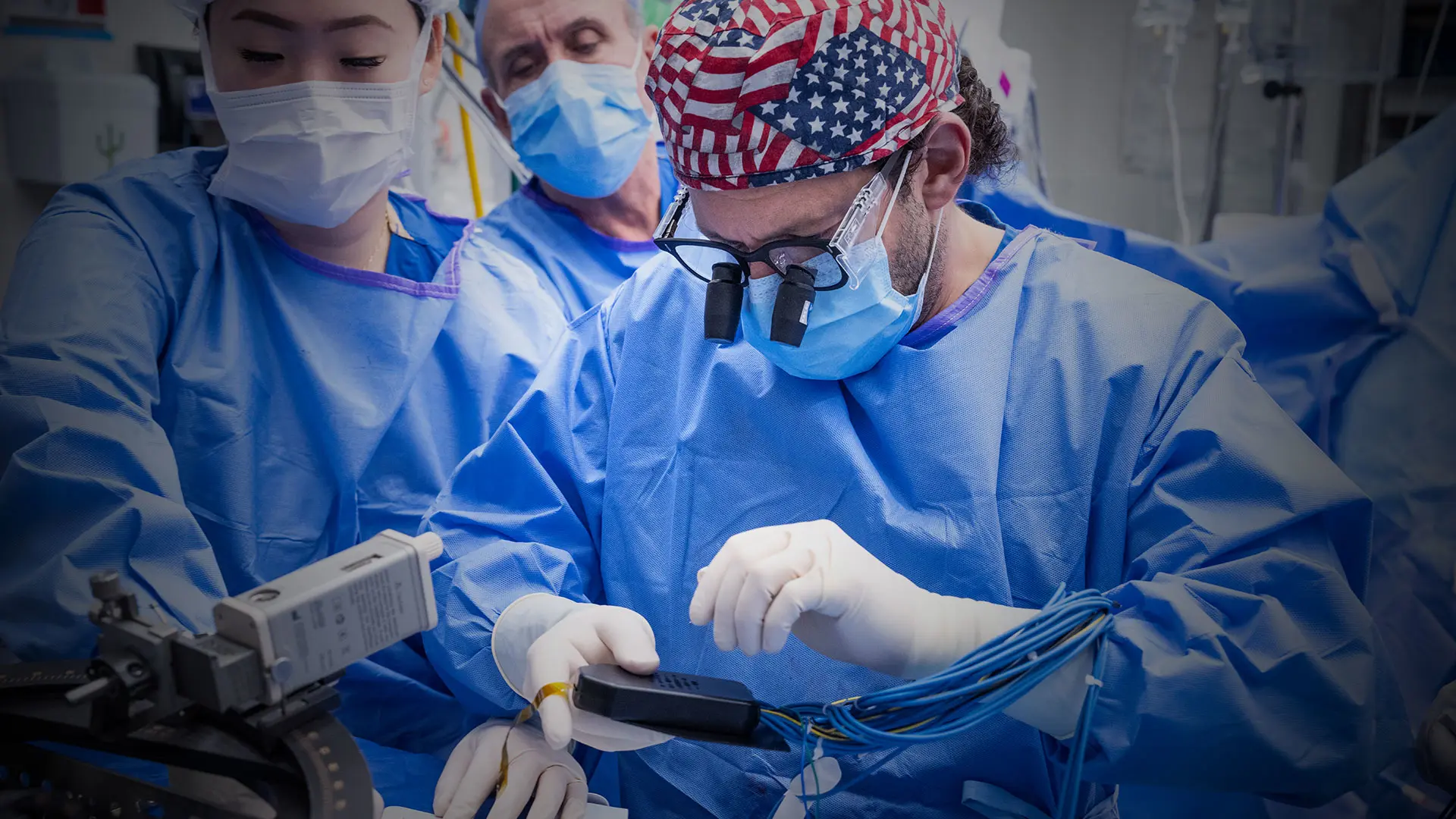
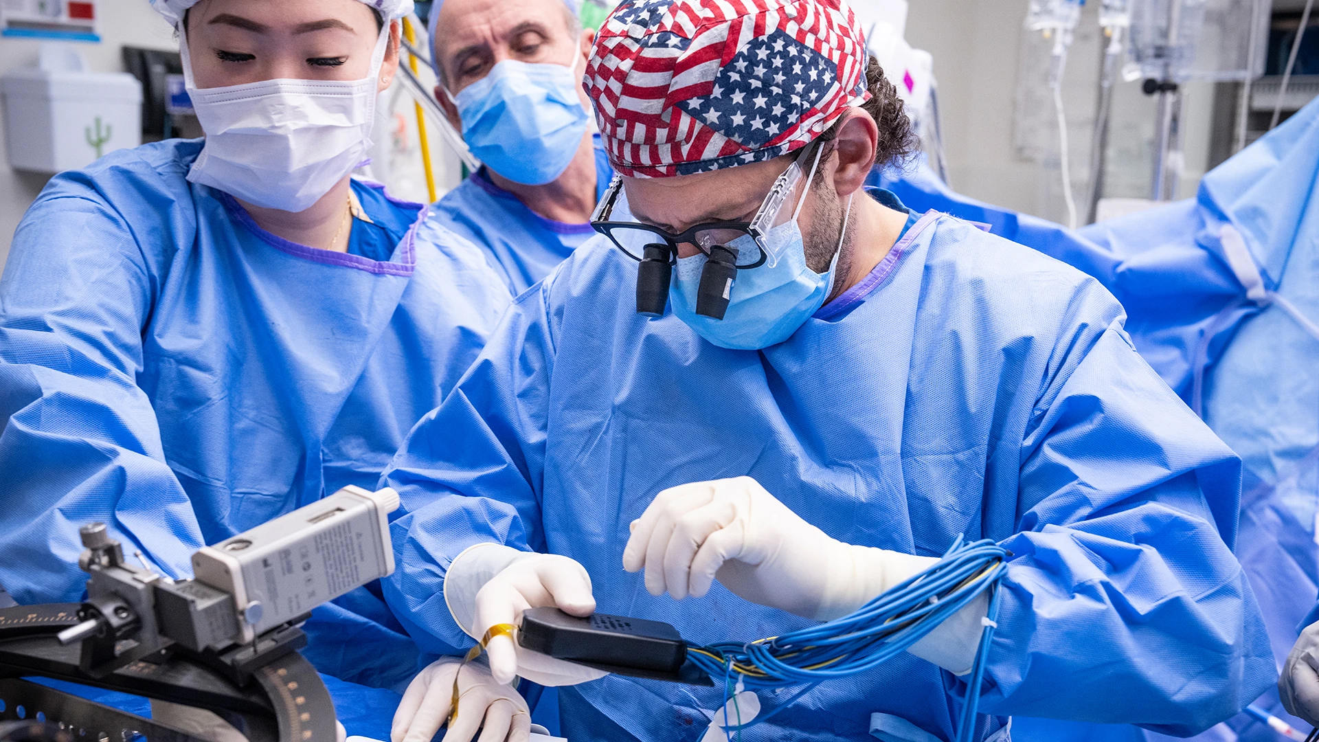
“Through the Living Brain Project at Mount Sinai, we developed a method to safely biopsy a small volume of brain tissue from a region of the prefrontal cortex during placement of the deep brain stimulation lead,” says Brian H. Kopell, MD, Director of the Center for Neuromodulation, co-lead of the Living Brain Project at Mount Sinai, and co-senior author of the paper. “The retrospective safety analysis clearly demonstrates there are safe and innovative ways of involving neurosurgical patients in research activities that have the potential to greatly advance science. We extend our deepest gratitude to the patients who have chosen to partner with us on this work.”
The research team analyzed acute adverse events, defined as infection, intracranial hemorrhage (ICH), and seizures, following 1,152 deep brain stimulation lead placements performed on 590 patients at a single hospital between 2013 and 2024. Prefrontal cortex biopsies were obtained in 652 procedures (“biopsy group”) and not obtained in 500 procedures (“non-biopsy group”). A computed tomography (CT) scan was performed within hours of each procedure and patient medical records were reviewed for acute ICH, seizure, and infection from the procedure date to 90 days later. A subset of patients was followed for approximately one year to assess cognitive outcomes.
“The retrospective safety analysis clearly demonstrates there are safe and innovative ways of involving neurosurgical patients in research activities that have the potential to greatly advance science. ”
— Brian H. Kopell, MD
No infections occurred in either group. No statistically significant differences in ICH rate or seizure rate were observed between groups (ICH rate: 1.7% biopsy group vs. 1.4% non- biopsy group; chi-square test p-value=0.88; seizure rate: 0.2% biopsy group vs. 0.4% non- biopsy group; p-value=0.82). No statistically significant associations were observed between number of biopsies and changes in cognitive health over time.
Brian H. Kopell, MD, monitoring electrodes of a Parkinson’s disease patient with Precision Neuroscience’s Layer 7 Cortical Interface.
“By using this technique, the Living Brain Project is already making several contributions to medical research, including a comprehensive characterization of living human brain biology, technology to study human brain circuits at the synapse level, technology to deliver gene therapy to the brain, and insights into the pathogenesis of brain disease,” says Alexander W. Charney, MD, PhD, Director of The Charles Bronfman Institute for Personalized Medicine, co-lead of the Living Brain Project, and co-senior author of the study. “Given the safety profile of the technique, we are eager for other institutions to follow the same methods and expand the scope of this work.”
Deep brain stimulation is an elective neurosurgical treatment for neurological and mental health illnesses. A common technique for safely implanting the DBS electrode involves cauterizing a small volume of prefrontal cortex prior to inserting the cannula.
“Given the safety profile of the technique, we are eager for other institutions to follow the same methods and expand the scope of this work.”
— Alexander W. Charney, MD, PhD
All the DBS procedures for the Living Brain Project were performed by a single neurosurgeon using standard stereotactic techniques. All procedures involved a standard frontal burr hole and stereotactic cannula placement, with cortical surface preparation differing only by the inclusion of a prefrontal cortex biopsy (obtained prior to cauterization and using a standard punch tool). Biopsy sizes were measured for 231 biopsies. The mean biopsy volume measured was equal to 40 mm3 and the median volume was equal to 30 mm3. The study authors note the tight biopsy sizes distributed around the mean demonstrate the Living Brain Project prefrontal cortex biopsy procedure can be highly standardized, reproducible, and precise.
“The innovative approach the Living Brain Project team carefully and meticulously developed has allowed analysis of living human brain tissue for the first time, transforming our capability to study the function of the human brain and providing new insights into the causes and treatment of brain diseases,” says Eric J. Nestler, MD, PhD, Anne and Joel Ehrenkranz Dean of the Icahn School of Medicine at Mount Sinai, and Executive Vice President and Chief Scientific Officer of the Mount Sinai Health System. “With more than a decade of data that demonstrate exemplary safety standards, the Living Brain Project is paving the way for other researchers to follow suit and help advance science beyond what has ever been possible before.”
Newer findings (Cell, January 22, 2026) from the Living Brain Project revealed how senescence processes involved in early brain development may also shape brain aging, providing new insight into how molecular signatures of cellular senescence that are present during development and aging mirror those associated with brain volume and cortical organization. The study, led by a team of Mount Sinai researchers, is the first to directly link senescence-related molecular networks in living human brain tissue to measurable differences in brain structure within the same individuals (see press release).
Two additional papers, published in 2025 and based on data from the Living Brain Project, uncovered evidence that brain tissue from living people has a distinct molecular character from tissue collected after death. The findings appeared in Molecular Psychiatry and PLOS ONE and call for a re-evaluation of how scientists study the human brain.
The Molecular Psychiatry study presents multiple lines of evidence that gene expression in postmortem human brain tissue may not always be an accurate representation of gene expression in living human brain tissue. Moreover, the study suggests that postmortem gene expression signatures of neurological and mental illnesses, as well as of normal phenotypes such as aging, may not always be accurate portrayals of those gene expression signatures in the living brain.
The study results published in PLOS ONE expand upon the Molecular Psychiatry study by focusing on RNA splicing and protein expression. Specifically, researchers learned the living brain is significantly different from the postmortem brain in terms of RNA splicing, intron usage, and protein expression. Study data revealed more than 60 percent of proteins and 95 percent of RNA types were differently expressed or processed in living versus postmortem tissue.
Led by Drs. Charney and Kopell, the Living Brain Project has enrolled more than 600 individuals undergoing elective DBS surgery for neurological and neuropsychiatric conditions including Parkinson’s disease, obsessive-compulsive disorder, essential tremor, dystonia, and depression.