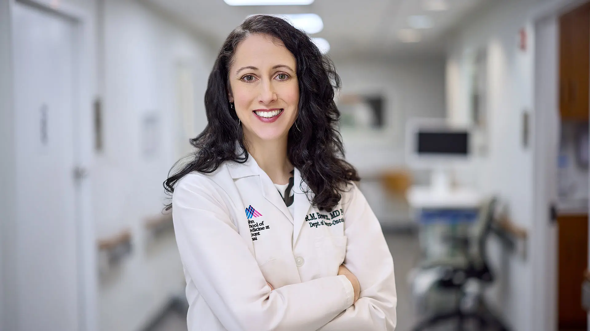
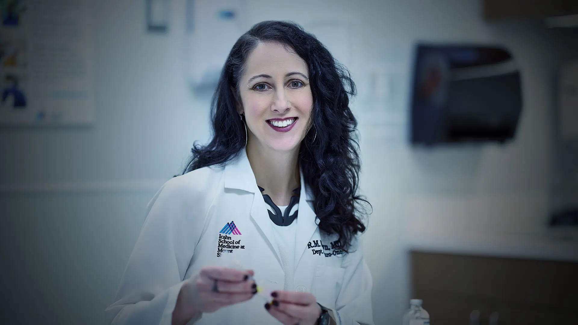
“These tumors dramatically affect patients’ well-being,” says Dr. Brown, Assistant Professor of Neurology, Neurosurgery, and Medicine (Hematology and Medical Oncology) at the Icahn School of Medicine at Mount Sinai. “I wanted to find a better way to help them.”

Rebecca M. Brown, MD, PhD
NF1 is a genetic condition characterized by the growth of tumors along peripheral nerves. While some patients develop cancerous tumors deeper within the body, the hallmark of NF1 is benign tumors located on or just under the skin. Those growths, or neurofibromas, can number in the thousands, causing itching and pain. The growths typically emerge in adolescence and often have devastating effects on a young person’s self-esteem and social and emotional well-being.
“Neurofibromas affect self-esteem and can lead to anxiety and depression. They contribute to relationship difficulties and even affect job prospects. Surgical resections could be widely adopted by other physicians in the neurology community.”
— Rebecca M. Brown, MD, PhD
Because the cutaneous growths are benign, most health insurance companies had not covered their removal. But after lobbying efforts by advocacy groups, insurance companies are now more likely to cover tumor removal, Dr. Brown says—an important development for these patients. “Neurofibromas affect self-esteem and can lead to anxiety and depression. They contribute to relationship difficulties and even affect job prospects,” she says. “Surgical resections could be widely adopted by other physicians in the neurology community.”
Neurofibroma Removal and Research
Currently, the only way to remove neurofibromas is with laser treatments or surgery. Lasers require anesthesia and lead to significant scarring and skin discoloration. Yet many physicians and patients have been wary of surgical resection because of frequent reports that the tumors quickly grew back. That has changed, thanks to a newly described surgical technique.
In 2019, Lu Le, MD, PhD, a dermatologist at UT Southwestern Medical School in Dallas, published a paper describing a protocol for resecting the tumors completely. Dr. Brown traveled to Texas to train with Dr. Le and now offers neurofibroma removal in her Mount Sinai clinic once a month. She can treat four or five patients a day, removing 15 to 18 tumors in each session. She prioritizes growths the patient finds most bothersome, whether due to discomfort or cosmetic concerns. “I’ve had a great success rate, with minimal scarring, zero infections, and very high patient satisfaction,” she says.
With patients’ permission, Dr. Brown sends resected tumors to Icahn Mount Sinai’s dermatology biobank. “This has gotten a lot of dermatology faculty interested in researching NF1,” says Dr. Brown, who is committed to improving lives for patients with NF1 and recently published a review paper in the journal Cancers describing the disorder’s dermatologic manifestations and emerging treatments.
She is also a primary investigator of a clinical trial testing a topical immunotherapy treatment for neurofibromas. In other work, she’s studying how gene expression differs from cell to cell within a neurofibroma and whether cells located on the skin behave differently from those deeper in the body. “If we’re going to target these tumors with medication, it’s important to understand the differences in cell behavior throughout the neurofibroma,” she says.
A Broader Role for Neurology in NF1
NF1 doesn’t have a natural home among specialties. Some patients receive care from neurologists, others from pediatricians, primary care doctors, or dermatologists. Yet even many dermatologists are not familiar with the protocol to surgically remove neurofibromas, Dr. Brown says. She maintains that neurologists—including neuro-oncologists like herself—are well suited to caring for patients with NF1. “When patients have such complex medical needs, having one physician who can address most or all of their needs improves continuity of care,” she says.
Youth diagnosed with NF1 are also at increased risk of autism spectrum disorder, attention-deficit/hyperactivity disorder, and learning disabilities. They may also experience skeletal abnormalities such as scoliosis and spinal tumors that contribute to severe chronic back pain. Treating these patients often includes helping them manage pain and the mental health effects of the disease, Dr. Brown says. Neurologists and neuro-oncologists can also follow patients to screen for the deeper tumors that may become cancerous, she adds.
Physicians in nonsurgical fields may hesitate to learn surgical resection of neurofibromas. But the procedure is not difficult to learn, Dr. Brown says. “Most of the neurofibromas I remove are a centimeter or less in size and require just one stitch,” she adds.
Dr. Brown welcomes physicians interested in training with her to learn the resection technique. “This is something that all neurologists who see NF1 patients could do to improve outcomes and satisfaction among our patients,” she says.
