Mount Sinai’s Department of Rehabilitation and Human Performance will lead the study, working closely with the Department of Neurosurgery. The first implantation in the United States of the Stentrode device, developed by Synchron, Inc., is expected to take place at Mount Sinai in the coming months.
BCI technologies aim to address failed transmission from the cerebral cortex. Surgically implanted neuroprostheses create a direct link from the brain to computers. For example, data from cortical surface electrodes have been used to produce natural speech, and using AI algorithms, to decode handwriting translated to texting. One such invention is the Stentrode, an endovascular stent and brain–machine interface already implanted in four humans and helping to translate their thoughts into digital output.
A recent publication in the Journal of NeuroInterventional Surgery demonstrated that two Australian patients implanted with the Stentrode learned to control texting and typing through direct thought. After training on mouse-clicking, they used the system unsupervised in their homes to send text messages, shop online, and manage their finances.
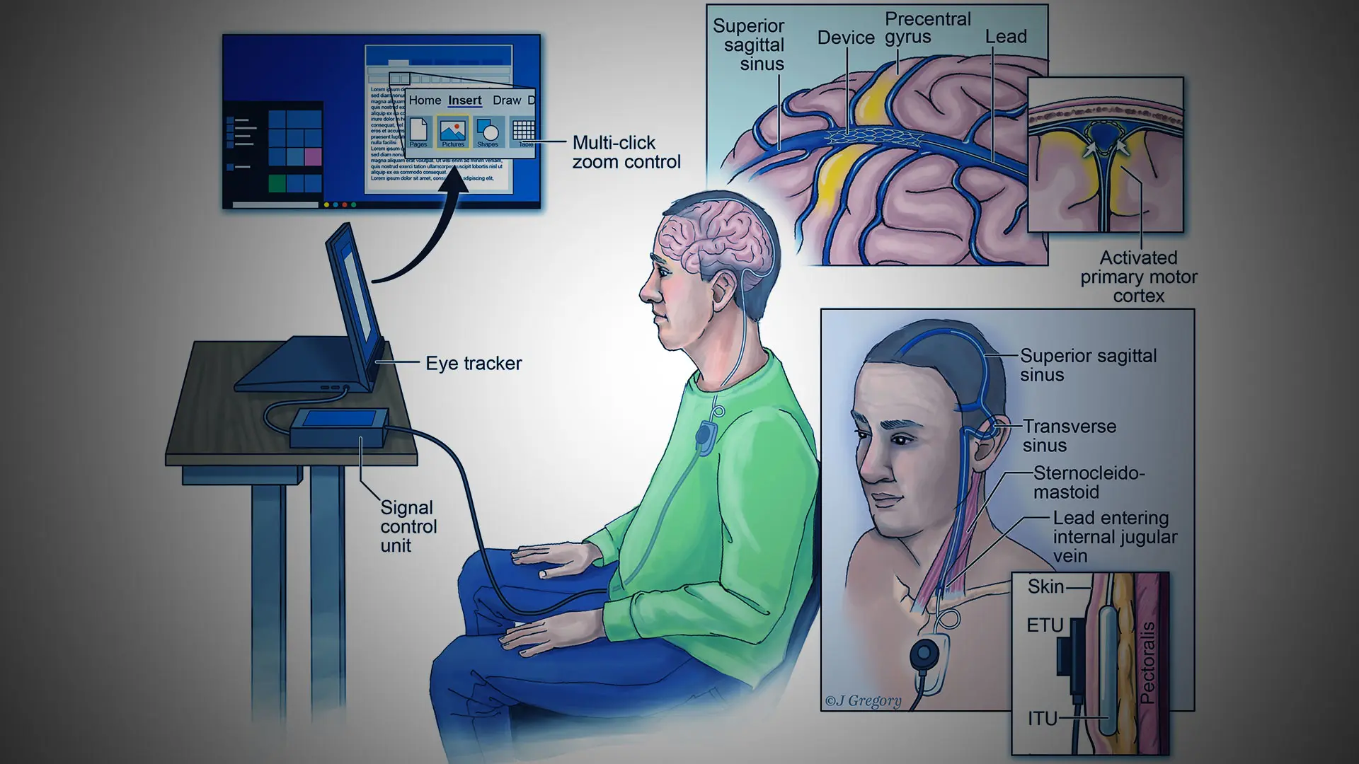
Endovascular motor neuroprosthesis system
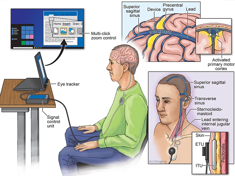
Above illustration: The internal and external system components in a participant with flaccid upper limb paralysis due to motor neurone disease are demonstrated. The device was implanted within the superior sagittal sinus, immediately adjacent to the precentral gyrus. The highlighted yellow region in the brain depicts the activation of primary motor cortex that occurs with attempted limb movement. The transmission lead, exiting the internal jugular vein between the heads of sternocleidomastoid, was tunneled subcutaneously and connected to the internal telemetry unit (ITU) placed within a subclavicular pocket. The external telemetry unit (ETU) inductively powers the ITU and receives the electrocorticography signal via infrared light transmission. The signal is sent to a tablet computer via a signal control unit and translated into multiple-click actions by the custom decoder, including a zoom function and single-click command. Multiple command control was combined with eye-tracking to enable general operation of Windows 10.
Shahram Majidi, MD, Assistant Professor of Neurosurgery, Diagnostic, Molecular and Interventional Radiology, and Neurology at the Icahn School of Medicine at Mount Sinai, will implant the devices for Mount Sinai.