Created in the research lab of Leif Havton, MD, PhD, Professor of Neurology, and Neuroscience, at the Icahn School of Medicine at Mount Sinai, the tool—known as shape-adjusted ellipse (SAE) approach—provides an innovative way of improving the accuracy of identifying and classifying nerve fiber types needed to generate ground truth data for both light microscopy (LM) and electron microscopy (EM) studies.
Tool improves accuracy of identifying, classifying nerve fiber types.
“When we have accurate measurements of the size and degree of myelination of nerve fiber types, we can better predict physiologic properties, such as how fast the nerve fiber impulses travel and how easy or difficult it is to activate them,” says Dr. Havton. “And, from that ground truth data, scientists and engineers can create better models, make better predictions, and develop vital new treatment devices for targeting select fiber types during neuromodulation by electrical stimulation.”
“When we have accurate measurements of the size and degree of myelination of nerve fiber types, we can better predict physiologic properties, such as how fast the nerve fiber impulses travel and how easy or difficult it is to activate them.”
– Leif Havton, MD, PhD
SAE could play a particularly important role in the rapidly emerging field of neuromodulation, which provides electrical stimulation to the brain or peripheral nerves through the activation of certain fiber systems to modulate abnormal neural pathways. Neuromodulation could result, for example, in new approaches for treating such neuro-urological conditions as overactive bladder syndrome, as well as pathologies as diverse as hypertension, gastrointestinal disorders, and inflammatory diseases.
The SAE approach is already a highly productive tool in the lab...
...and we have made it accessible for any researcher to use.
The SAE approach for assessing nerve fibers is already a highly productive tool in Dr. Havton’s lab as part of its ongoing neuromodulation research, which includes groundbreaking work recently published in Scientific Reports. The study shares links to the SAE approach tool that researchers can access and use.
Dr. Havton and his research team at Mount Sinai are also members of, and funded by, two prominent research consortia: the National Institutes of Health SPARC (Stimulating Peripheral Activity to Relieve Conditions) program, and the Dr. Miriam and Sheldon G. Adelson Medical Research Foundation. Both consortia are focused on supporting collaborative research work that has the potential to dramatically change biomedicine in the decade ahead.
Scientific tools to quantify the sizes and classification of fibers and myelination have changed very little over the past century. The SAE approach for determining physiologic phenotypes and developing improved theoretical neuromodulation models has become a game-changer, made possible when significant advances in light and electron microscopy revealed new insights into the fine organization of the nervous system at high resolution, giving scientists innovative ways to overcome previous research obstacles.
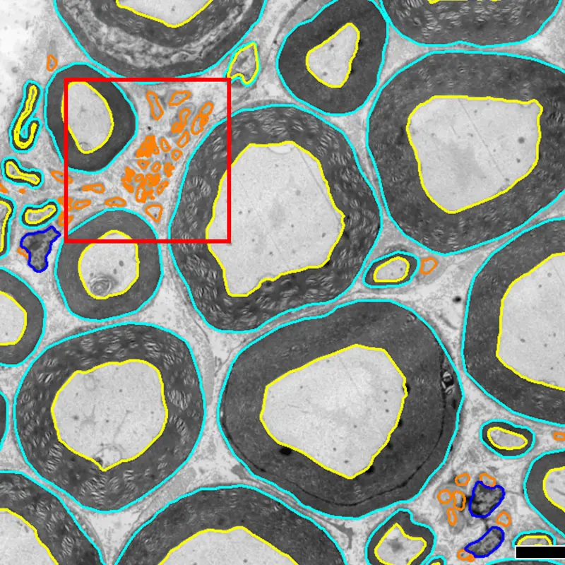
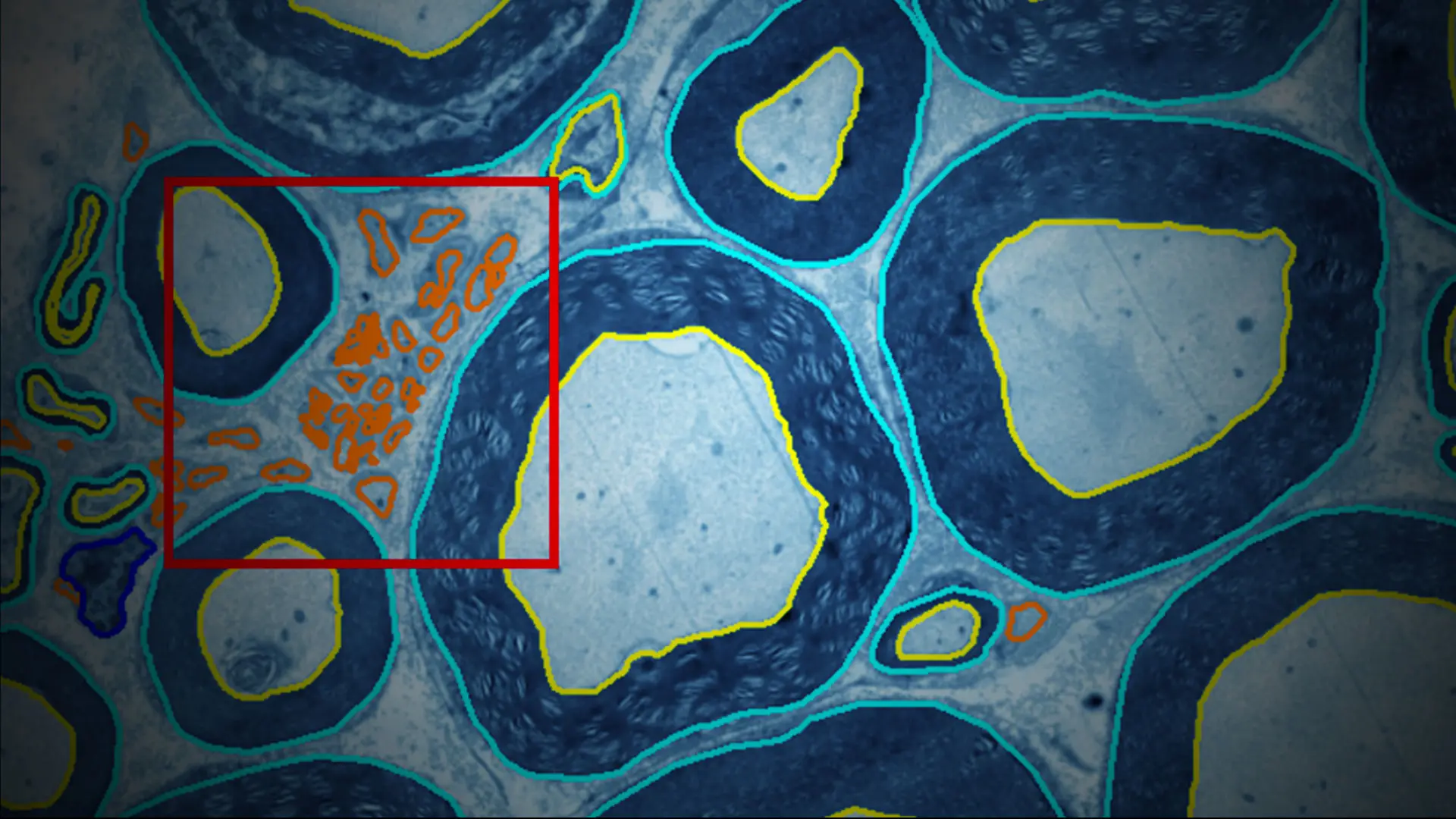
Transmission electron microscope images of lumbosacral ventral roots in rhesus macaques. Computer-supported analysis allows for segmentation of myelinated and unmyelinated axons. Boxed area shown at higher magnification in next image. Scale: 10 µm
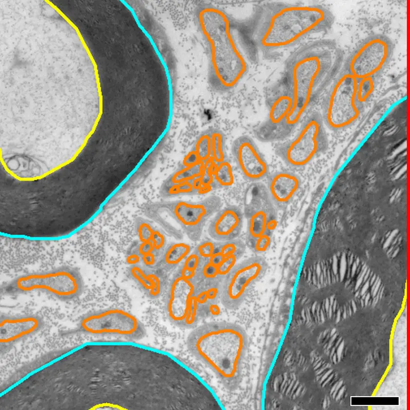
Outer and inner borders of myelinated axons shown in light blue and yellow, respectively; outline of unmyelinated axons is contoured in orange. Digital measurements of nerve fiber area and perimeter from each axon are used to determine a corrected value of fiber size based on SAE approach. Scale: 1 µm
Those revelations allowed researchers, for example, to resolve these perplexing challenges:
Segmentation of axons in light and electron micrographs allows for quantitative high-resolution analysis of nerve fiber tissues—but varied axonal angles result in an overestimation of fiber sizes. The SAE approach today corrects for size and myelination of individual nerve fibers for light and electron microscopy by compensating for the shape and dispersion angle of individual nerve fibers.
The correction for fiber dispersion angle markedly reduces the number of outliers and data scatter, allowing for enhanced anatomical mapping as well as improved predictions of functional properties in translational studies of neural repair and nerve regeneration.
The SAE approach was validated by light and electron microscopy against the traditional methods of determining nerve fiber size and myelination—which had resulted in significant overestimations of fiber diameters.
“We’re receiving more and more inquiries from researchers across the United States and abroad.”
– Leif Havton, MD, PhD
The new precision tool is the result of a unique merger within Dr. Havton’s lab of the mathematical and engineering skills of Petra Bartmeyer, PhD, and the neuroscience and light and electron microscopy expertise of Natalia Biscola, PhD, both postdoctoral researchers in the Department of Neurology at Mount Sinai.
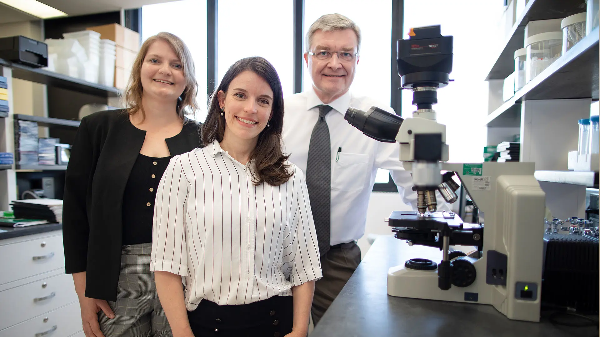
Leif Havton, MD, PhD, with postdoctoral fellows Natalia Biscola, PhD, center, and Petra Bartmeyer, PhD
“We’re extremely pleased with the increased accuracy and interpretability of the data that have been generated through our team’s tireless work,” says Dr. Havton. “We’re receiving more and more inquiries from researchers across the United States and abroad, and we are proud that we’re now able to share this tool with the entire neuroscience community.”