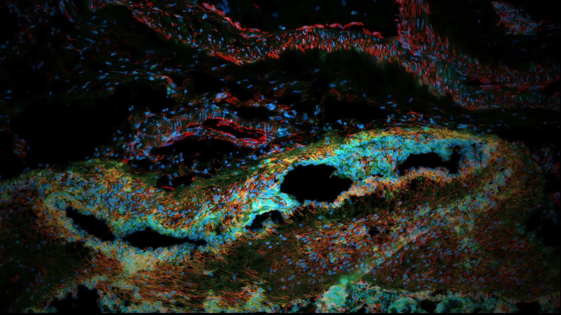
“I have found it particularly eye-catching how vessels of the choroid plexus show elongated nuclei, creating an appearance like flowing water,” says Sarah Murphy, an Associate Researcher at the Center for Disease Neurogenomics, who uses the latest "spatial omics" tools to study common neuropsychiatric and neurodegenerative disorders. “The dendrite branching of the neuronal cells also seen in this image show a similar display of flow. This is all conceptually thought-provoking when you think of how information is likely being passed among these cells, following the flow of their morphology.”
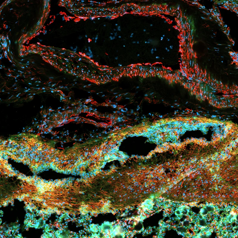
This image was captured on the Xenium Analyzer from 10X Genomics, an innovative imaging platform used to generate high-resolution single-cell spatial transcriptomic datasets.
The human choroid plexus is responsible for producing the cerebrospinal fluid that normally bathes and cushions the brain and that is also associated with several disorders. In her composite image, blue corresponds to DAPI to indicate nuclei, green corresponds to ATP1A1, CD45, and E-cadherin to mark cell membrane boundaries. Yellow indicates 18S rRNA to mark cytoplasmic RNA, and red corresponds to alphaSMA and vimentin to mark intracellular proteins.
“When generated using ex vivo human brain specimens, high-resolution single-cell and spatially resolved datasets deepen our understanding of the molecular basis of neuropsychiatric and neurodegenerative disorders,” she says. “Case vs. control studies applied to disease-relevant regions of the brain hold the potential to move us closer to developing and enhancing precision medicine across a range of debilitating disorders.”
CREDIT:
Sarah Murphy, Associate Researcher, Center for Disease Neurogenomics
Lab of Panagiotis (Panos) Roussos, MD, PhD
Professor of Psychiatry, and Genetics and Genomic Sciences, Director of the Center for Disease Neurogenomics, and a member of The Friedman Brain Institute