Stress disrupts brain-body communication through top-down (brain-to-body) and bottom-up (body-to-brain) mechanisms, influencing processes such as interoception and energy homeostasis. Interoception, the brain's ability to sense and interpret internal bodily signals, such as heart rate, respiration, or energy metabolism, supports emotional regulation, decision-making, and stress responses, while maintaining homeostasis by addressing bodily needs.
The autonomic nervous system, comprising the sympathetic (SNS) and parasympathetic (PNS) branches, governs emotional and physiological responses to stress. The SNS drives fight-or-flight-or-freeze responses to stressors, increasing heart rate and respiration while suppressing digestion. The PNS promotes rest-and-digest states, counterbalancing stress responses. While SNS activity has been extensively studied, less is understood about how the PNS counteracts defensive reactions. Understanding the mechanisms of the PNS is crucial for addressing conditions such as anxiety or panic disorders that are highly comorbid with metabolic, immune, and cardiovascular diseases.
The vagus nerve, a key regulator of the PNS, serves as a critical pathway for interoceptive signals, transmitting information from the heart, lungs, and other organs to the brain. At the brainstem level, the nucleus of the solitary tract (NTS) acts as the central hub for vagal inputs, processing visceral information and regulating autonomic, emotional, and physiological states. The NTS relays these signals to the insular cortex, which integrates sensory information—such as heart rate, breathing, and hunger—to shape conscious perceptions of bodily states and guide emotional and behavioral responses.
The lab of Abha Karki Rajbhandari, PhD, focuses on understanding how the brain, vagus nerve, and body interact, particularly in relation to stress and energy metabolism.
Disruptions in communication along the pathway from the vagus nerve to the insular cortex are linked to conditions such as depression, post-traumatic stress disorder (PTSD), cardiovascular disorders, and metabolic issues. Conversely, better communication in this circuitry supports improved emotional regulation and stress resilience. The posterior insular cortex is closely associated with the brain's fear circuitry, connecting to the amygdala, where heightened activity is correlated with defensive reactions (see Figure).
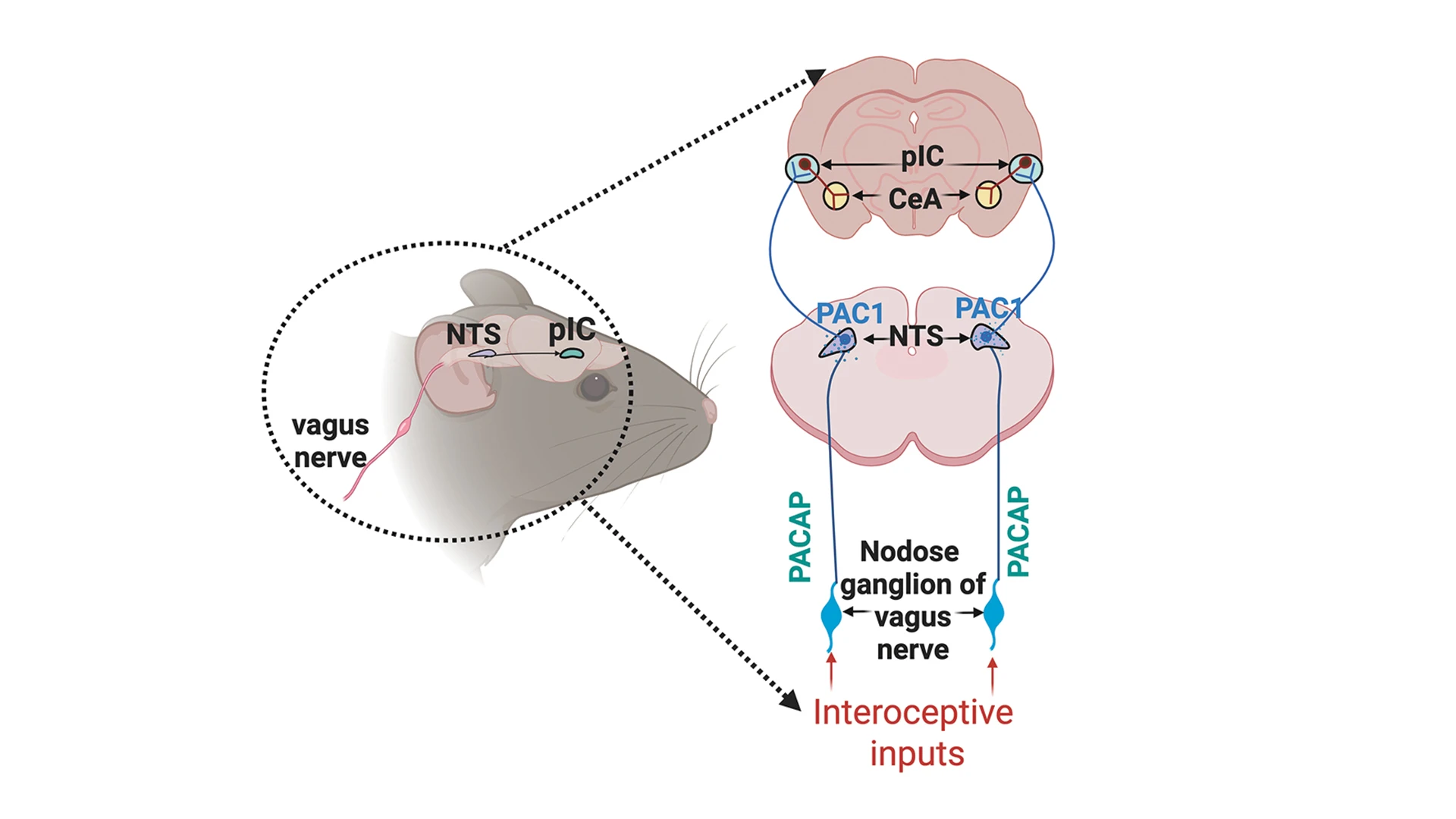
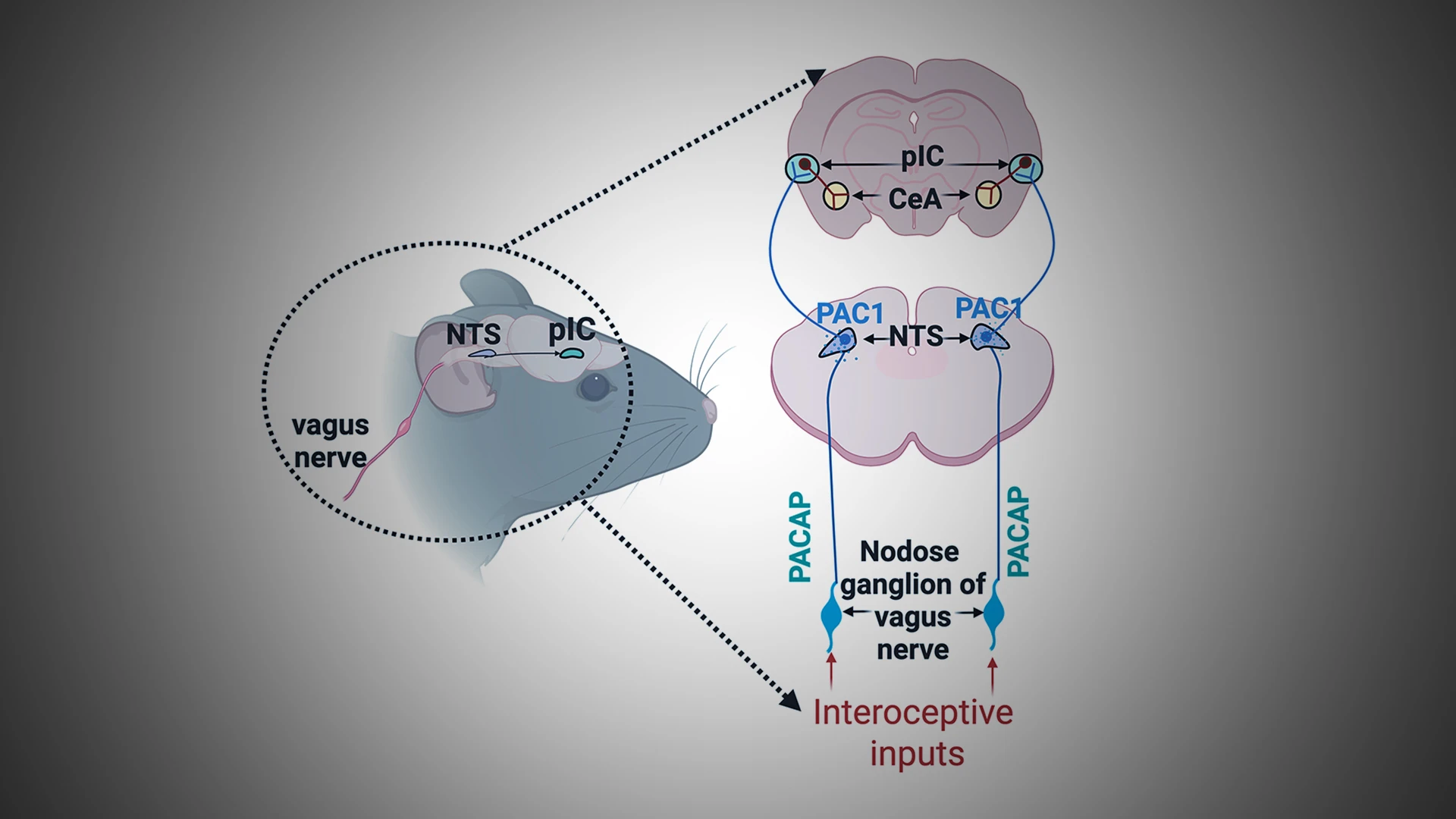
Figure. Simplified Model of Brain Regions Involved in Interoceptive Pathways Mediating Stress Responses
This simplified model highlights the brain regions involved in interoceptive pathways that mediate the body-to-brain communication during stress responses. It illustrates how visceral inputs from organs such as the heart and lungs travel to the brainstem's NTS and then to the insular cortex (IC) in the forebrain, ultimately influencing the central amygdala (CeA). Dysregulation at any point in this pathway under stress can lead to heightened fight-or-flight-or-freeze responses, contributing to anxiety disorders often comorbid with cardiovascular and metabolic diseases. Neuropeptides such as PACAP, expressed in the vagus nerve’s nodose ganglion, play a key role in regulating these interoceptive pathways. Understanding the functions of these pathways and their components provides opportunities to develop novel therapeutic approaches for a growing range of stress-related conditions.
Activation of the vagus nerve promotes relaxation and recovery, a function often referred to as the "vagal brake." Chronic stress can weaken vagal tone, prolonging fight-or-flight-or freeze responses and exacerbating mental, cardiovascular, and metabolic conditions. Practices such as deep breathing, meditation, and vagus nerve stimulation enhance parasympathetic activity, improving interoceptive awareness and emotional resilience. Hence, precise manipulation of the vagus nerve offers promising new therapeutic approaches for a range of conditions.
The Karki Lab focuses on investigating brain-body interactions under stress by delineating the role of parasympathetic pathways in regulating stress-related behaviors, cardiorespiratory functions, and energy homeostasis.
Studies suggest that multiple neuromodulators within the vagus nerve regulate specific aspects of behavioral and emotional responses. The Karki lab focuses on investigating brain-body interactions under stress by delineating the role of parasympathetic pathways in regulating stress-related behaviors, cardiorespiratory functions, and energy homeostasis. This interdisciplinary approach merges neurobiology with cardiology and metabolism, advances our understanding of stress biology, and uncovers new avenues for addressing the effects of stressors.
Using mouse models, the lab has discovered specific neuropeptidergic pathways from the vagus nerve's nodose ganglion to the NTS that involve the neuropeptide PACAP.
Chemogenetic activation of this pathway in the nodose ganglion of the vagus nerve lowers fear levels and improves heart rate variability worsened by stress-enhanced fear learning (SEFL), a behavioral paradigm that mimics aspects of PTSD-like fear (seeJournal of Visualized Experiments and Neuroscience Biobehavioral Reviews.) Conversely, experimentally inhibiting the insular cortex (for example, with chemogenetic approaches) along with nodose PACAP stimulation decreases fear levels, highlighting the significance of this pathway. The lab has also shown that SEFL paired with nodose PACAP stimulation increases calcium activity in the insular cortex, as observed via fiber photometry, compared to SEFL alone. These findings suggest that nodose signaling to the insular cortex and amygdala communicates that "something significant has occurred," guiding state-dependent fear responses. Thus, our cell- and gene-specific investigation of the vagus nerve-to-insular cortex pathway in interoceptive processes could enable targeted interventions for stress-related conditions.
As stress-related conditions continue to rise in society, emerging fields focusing on interoception and energy metabolism are transforming our understanding of human health. The vagal system, linked to the NTS, also expresses glucagon-like peptide-1 (GLP-1) receptors, which play a broader role in regulating behavioral, autonomic, and neuroendocrine responses to psychogenic stressors. Studies conducted in the lab of Paul J. Kenny, PhD, at Mount Sinai also show that GLP-1 signaling through NTS influences physiological and behavioral responses. (See related article.)
At Mount Sinai, researchers are advancing therapeutic approaches by integrating insights into brain-body communication to develop innovative treatments and improve health outcomes. For example, our collaborative efforts with scientist Filip Swirski, PhD, Director of the Cardiovascular Research Institute, are enhancing our research into the intricate relationship between the brain and heart. We believe these intricate and mechanistic studies focused on neuroscience, cardiology, and metabolism will someday lead to preventive and therapeutic strategies, and possibly uncover biomarkers and mechanisms of biofeedback for stress-related conditions that are often comorbid with cardiovascular and metabolic diseases.
Featured

Abha Karki Rajbhandari, PhD
Assistant Professor of Psychiatry, and Neuroscience

Filip Swirski, PhD
The Arthur and Janet C. Ross Professor of Medicine (Cardiology), Professor of Diagnostic, Molecular and Interventional Radiology, and Director of the Cardiovascular Research Institute