MRI is also an essential tool in neuroscience, allowing for the study of brain function and metabolism in addition to structure in humans and in animals, including rodent and nonhuman primate models.
Brain imaging research at the Icahn School of Medicine at Mount Sinai is led by the Advanced Neuroimaging Research Program (ANRP), a collaborative effort between The Friedman Brain Institute and the BioMedical Engineering and Imaging Institute (BMEII), which aims to improve the performance of all neuroimaging modalities. Priti Balchandani, PhD, serves as Director of the ANRP and Associate Director of the BMEII.
Icahn Mount Sinai is one of the few institutions that has invested in a 7-Tesla (7-T) human MRI scanner, which produces high-resolution images to visualize and measure previously undetectable changes in the brain.
This includes the use of functional MRI to study brain function, diffusion MRI to study brain networks and white matter tracts, and MR spectroscopic imaging for the study of metabolism. These methods are used clinically to diagnose and plan treatment for numerous conditions, including epilepsy, brain tumors, multiple sclerosis, and brain and spinal cord injury. These methods are also used to gain insight into brain abnormalities that underlie all major psychiatric disorders, including schizophrenia, bipolar disorder, depression, and drug addiction.
Icahn Mount Sinai is also one of the few institutions that has invested in a 7-Tesla (7-T) human MRI scanner, which produces high-resolution images to visualize and measure previously undetectable changes in the brain. MRI at 7-T is completely noninvasive and well tolerated by patients. This advanced imaging technology captures more subtle brain abnormalities, enabling earlier detection and treatment of diseases that are not detected through standard imaging. Using 7-T, Mount Sinai can perform deeper analysis of smaller structures in the brain—such as specific hippocampal subfields and brainstem monoaminergic nuclei—that are involved in multiple disease etiologies and not discernable with standard approaches.
Multimodal MRI exams are composed of several MRI pulse sequences, each imparting an important aspect of tissue structure, function, vascularity, and metabolism. MRI at ultrahigh magnetic fields produces structural images with exquisite resolution, well beyond clinically available MRI and can elucidate more subtle abnormalities in the brain structure, function, and connectivity.

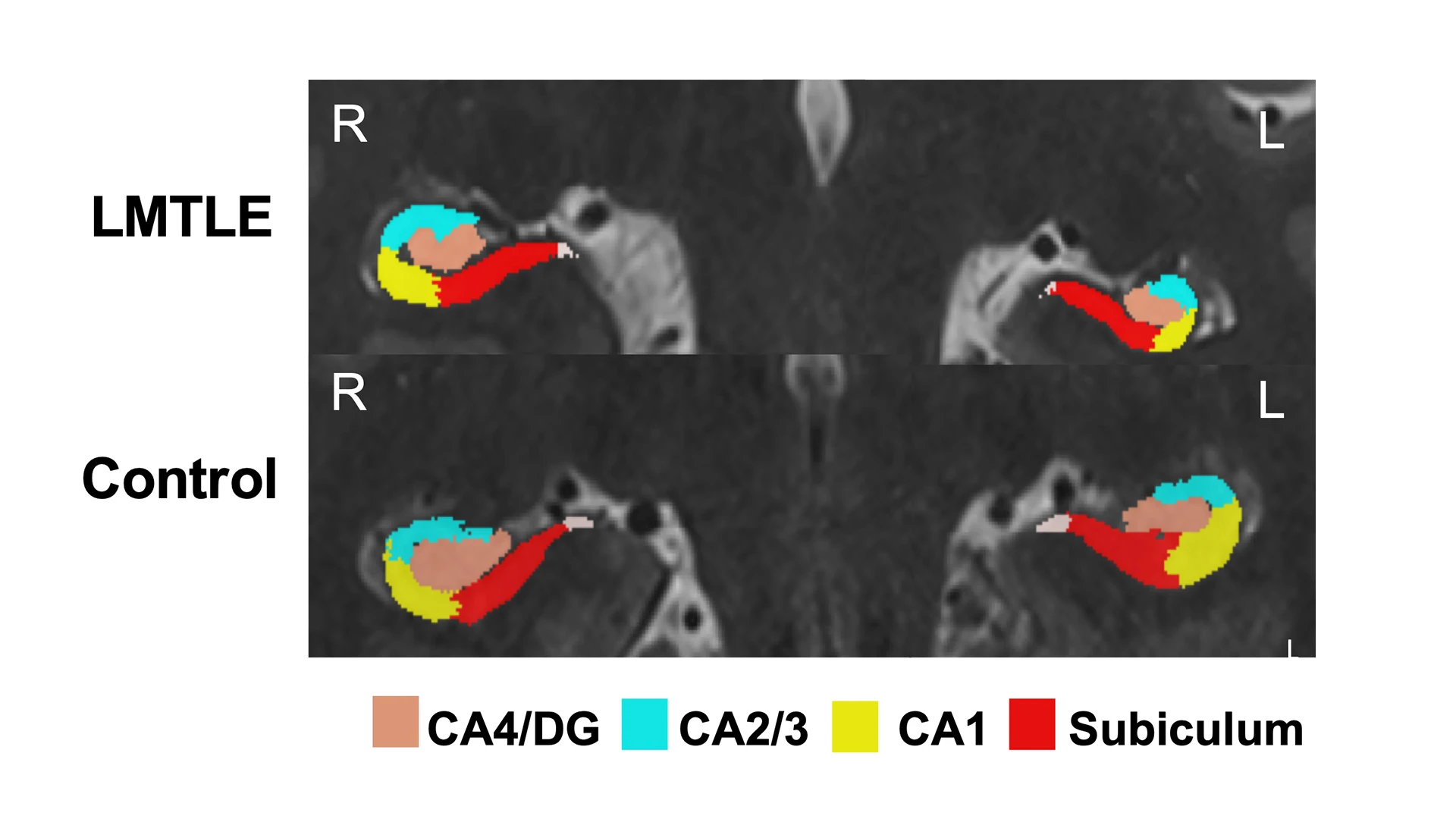
Figure 1. Hippocampal subfield segmentation for epilepsy patient and control. This compares hippocampal subfield volumes in a mesial temporal lobe epilepsy patient and a healthy control and demonstrates the level of detail achievable through high-resolution structural images.

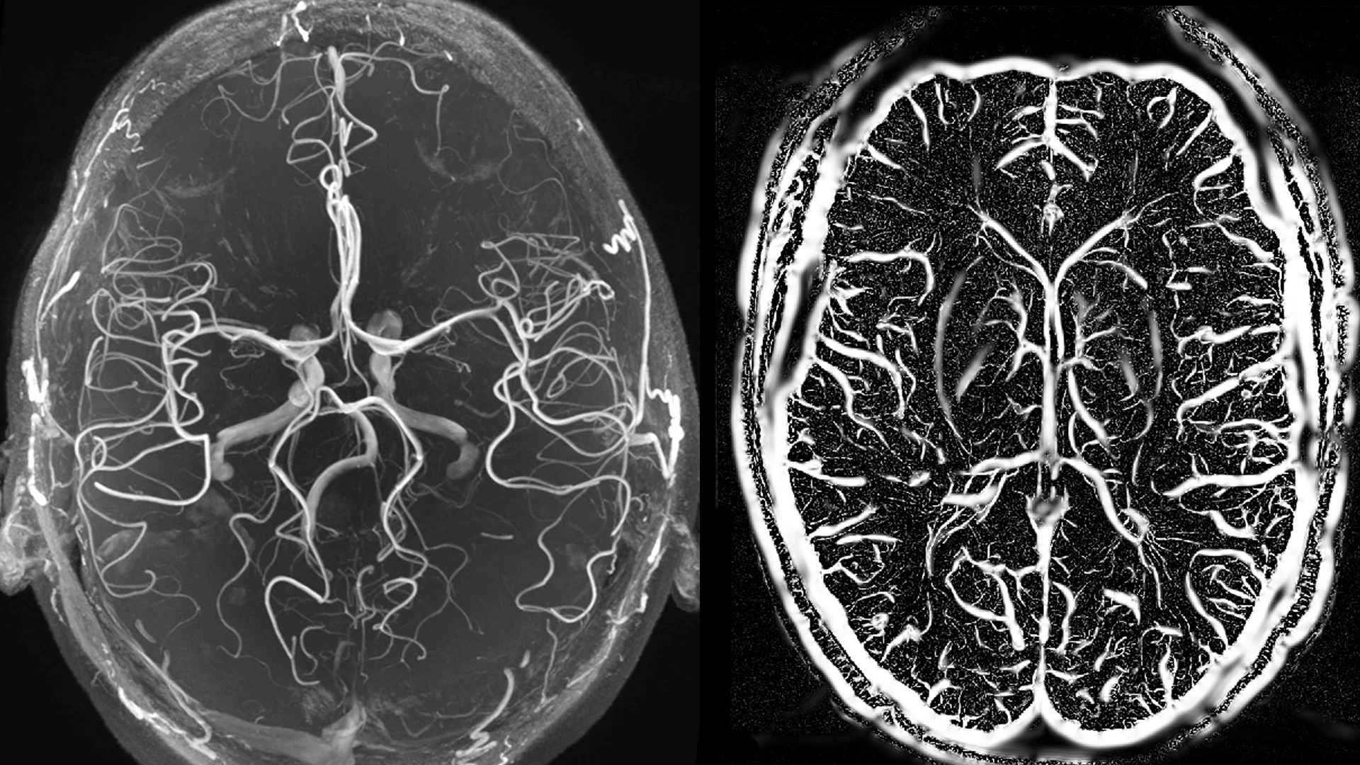
Figure 2. 7-T time of flight (TOF) image showing arteries (left) and SWI segmentation showing veins (right). This depicts 7-T TOF imaging and susceptibility-weighted imaging (SWI) allow visualization of vascular abnormalities that may underlie structural lesions.
Resting state functional MRI (rs-fMRI) at 7-T can provide highly resolved activation maps that can detect abnormal brain function in disease but also act as a powerful neuroscience tool to study cognition and behavior. Magnetic resonance spectroscopic imaging (MRSI) captures signals from brain metabolites, such as N-acetyl aspartate (NAA) and creatine (Cre), to provide information about neuronal integrity and total energy metabolism. Overall, multimodal imaging at ultrahigh fields is now being used to reveal underlying mechanisms of diseases such as non-lesional epilepsy, Alzheimer’s disease, and idiopathic neuropathic pain through novel detection of abnormalities in brain regions and circuits that are integral to disease pathophysiology.

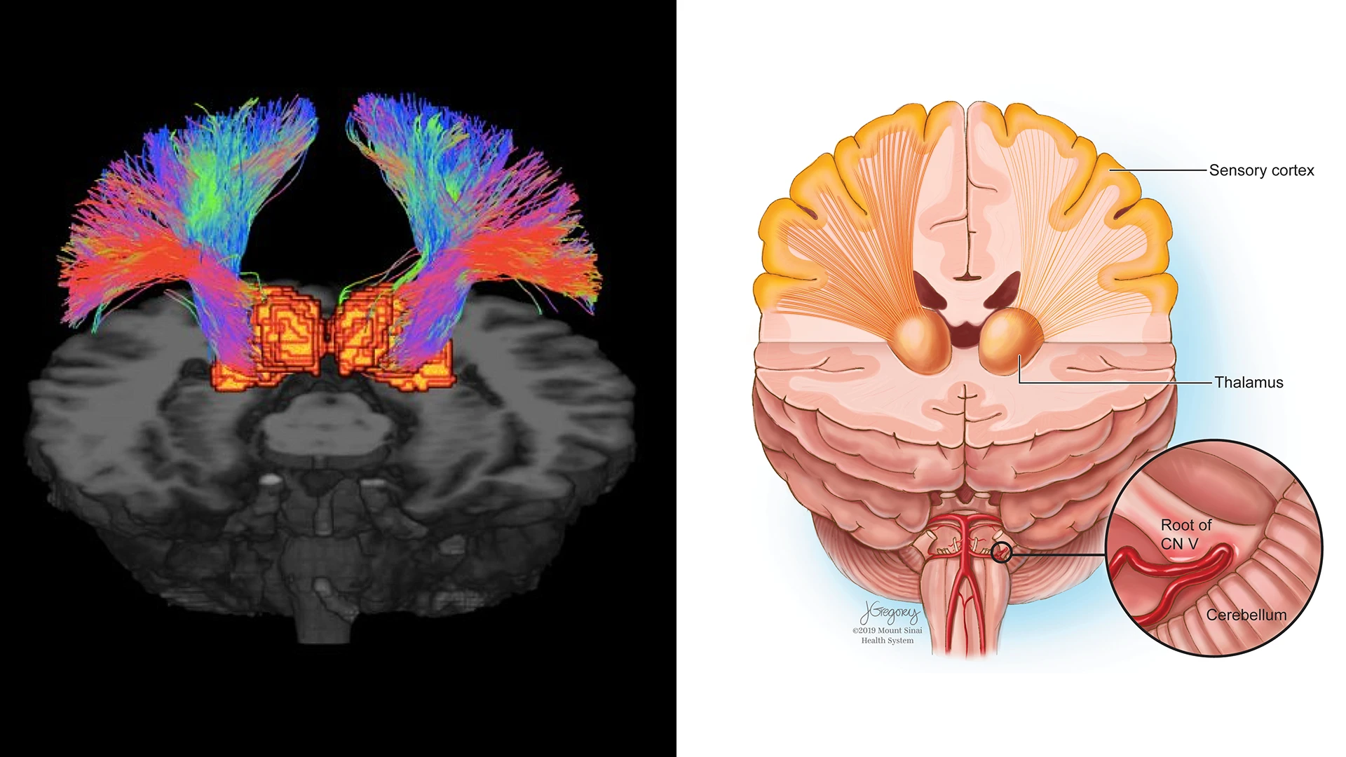
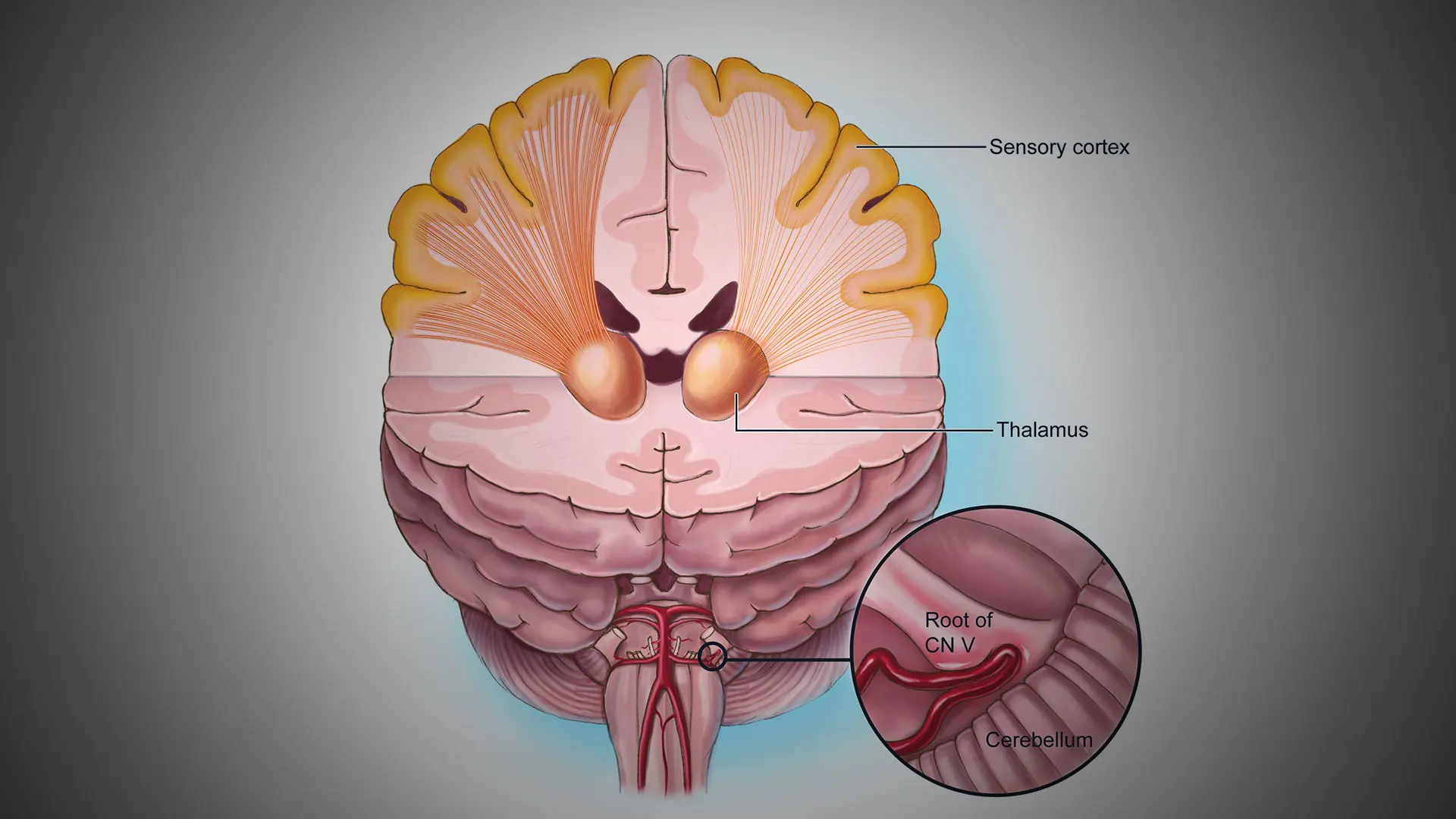
Figure 3. Probabilistic tractography of the thalamic-somatosensory tracts. Trigeminal neuralgia (TN) patients exhibited reduced integrity of these tracts on the ipsilateral side when compared to controls. These depict the white matter tracts connecting the thalamus to the somatosensory cortex in TN, a debilitating neuropathic pain condition that is often misdiagnosed due to lack of etiological understanding. 7-T diffusion weighted MRI (dMRI) is ideal for detecting changes in microstructure and integrity of white matter tracts and nerve fibers.
Beyond detection of qualitative abnormalities, advanced image analysis methods quantify information such as volumes of important brain structures, microstructural integrity of brain tissue, white matter connectivity of brain networks, and metabolite ratios for important brain regions. These quantitative metrics are used to improve detection of abnormal tissue structure and function and aid in more targeted, biologically driven interventions. Multiparametric quantitative approaches may also be used to calculate abnormality indices that can assist clinicians in decision-making. Once imaging data are quantified, the findings are combined with genetic and clinical data to power more personalized clinical decision-making and improve overall understanding of the brain in health and disease.
Featured

Priti Balchandani, PhD
Professor of Diagnostic, Molecular and Interventional Radiology, Neuroscience, and Psychiatry; Director, Advanced Neuroimaging Research Program; and Associate Director, BioMedical Engineering and Imaging Institute
