Investigating the mechanisms that drive phenomena such as scarring and wound regeneration in the vocal folds is of interest to Benjamin Laitman, MD, PhD. But accessing healthy tissue for analysis can often be difficult.
“It is a very delicate structure, so you don’t want to biopsy it and risk damage,” says Dr. Laitman, an otolaryngology resident at The Mount Sinai Hospital. “You typically do not remove any of it unless it is already diseased.”
Obtaining healthy vocal folds from cadavers, he adds, is also challenging because there are often delays in harvesting, and healthy tissue typically is not taken at autopsy. However, a new technology, combined with access to fresh healthy tissue, is helping Dr. Laitman gain insight into the vocal folds without having to rely on traditional methods.
“We are using tissue that would otherwise go to waste to conduct high-level genetic sequencing to understand which cells are present in the vocal folds,” he says.
Using Cell-by-Cell Sequencing Technology to Map the Vocal Folds in High Resolution
Using tissue resected from patients who have undergone pitch elevation surgery—tissue that would otherwise be discarded—Dr. Laitman is collaborating with the Genomics Core Facility at the Icahn School of Medicine at Mount Sinai to create a high-resolution map of the human vocal folds. He is achieving this goal with 10x Genomics’ single-cell RNA-seq (scRNA-seq) technology, which enables researchers to analyze transcriptomes—the full range of messenger RNA molecules expressed by an organism—on a cell-by-cell basis using microfluidic partitioning to capture individual cells.
“This technology gives us a much higher-resolution look at what is going on genetically as opposed to a bulk RNA sequencing approach,” he says. “Through cell-by-cell sequencing, we are developing a baseline map of the vocal folds that we can use as a basis for comparison when looking at diseased tissue, be that benign disease, cancer, or scarring.”
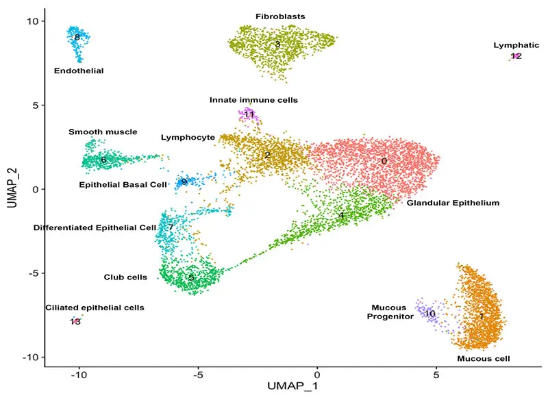
Single-cell RNA sequencing of human vocal folds. Uniform manifold approximation and projection (UMAP) plot of human vocal fold clusters.
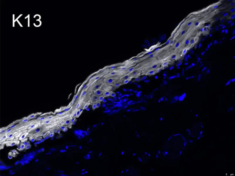
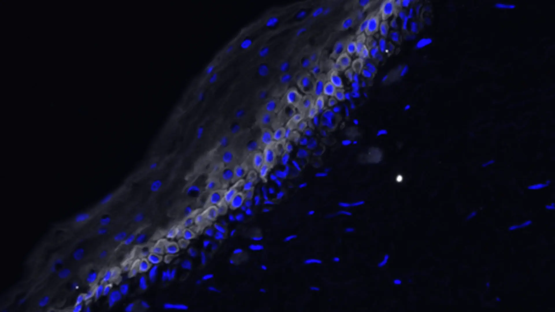
Laryngeal epithelial microarchitecture. Immunofluorescent image of human laryngeal epithelium Keratin 13 (K13) is expressed in more differentiated layers, while K14 is expressed in basal layers.
The effort, which began earlier this year with a grant from the American Laryngological Association, is one of the first-ever attempts to map the vocal folds using this technology. It is an undertaking that has gained urgency in the wake of the pandemic, which resulted in many patients being intubated for management of COVID-19-related acute respiratory failure, putting them at risk for developing life-altering complications.
“Intubation can impact everything from how the patient’s voice sounds to their ability to breathe and swallow,” Dr. Laitman says. “Even before the pandemic, there was a big push to understand the dynamics of wound healing and scar formation in the airways to address those risks. Although we have some understanding of the cells regulating that process, we need a more comprehensive picture.”
Advances in technology, such as single-cell sequencing, are delivering those insights, enabling Dr. Laitman to explore the types of cells that are present in the vocal folds to a degree that was not previously possible. More importantly, it creates opportunities for potentially co-opting those cells in ways that benefit the patient.
“For example, if we identify certain types of fibroblasts that are usually involved in scarring, we could possibly block those while potentially stimulating others that promote wound generation,” he says.
Mapping Disease-State Samples
But there is considerable work to do before such benefits can be realized. Dr. Laitman and the team have sequenced a set of five samples, effectively mapping out one region of the vocal folds. Any findings as to the drivers of wound regeneration or scarring in this region may not be applicable to the other regions of the vocal folds. Thus, the goal is to complete the atlas of the vocal folds so that Dr. Laitman can begin what he characterizes as the most interesting and important step in his research: mapping disease-state samples.
“The sequencing technology has advanced to the point where we can use previously banked, paraffin-embedded samples and not only achieve high-resolution single-cell data, but also spatial sequencing data where we can put the genes in context to map them out,” Dr. Laitman says. “We can then compare the healthy- and disease-state maps, zero in on the cells at play in the disease state, and see both the differences in the cells and the genetic pathways that are activated. That would facilitate the identification and development of therapeutic agents to address these differences.”
For now, Dr. Laitman has the foundation for what he believes will be a hypothesis-generating resource that will be invaluable for Mount Sinai and other academic medical centers. “I am really excited about what could emerge from the work we are doing,” he says.
Featured
Benjamin Laitman, MD, PhD
Otolaryngology Resident, The Mount Sinai Hospital