Mount Sinai dermatologists are accelerating their research efforts in the diagnosis of skin cancer through their leading-edge work with technologies like reflectance confocal microscopy, Vectra® total body photography, and, most recently, artificial intelligence (AI).
“Our ability to diagnose skin cancer accurately has the potential to become exponentially better,” says Jonas Adalsteinsson, MD, PhD, Assistant Professor of Dermatology at the Icahn School of Medicine at Mount Sinai, who is actively involved in that research. “We’re making steady progress, and I think the future is very bright for our field.
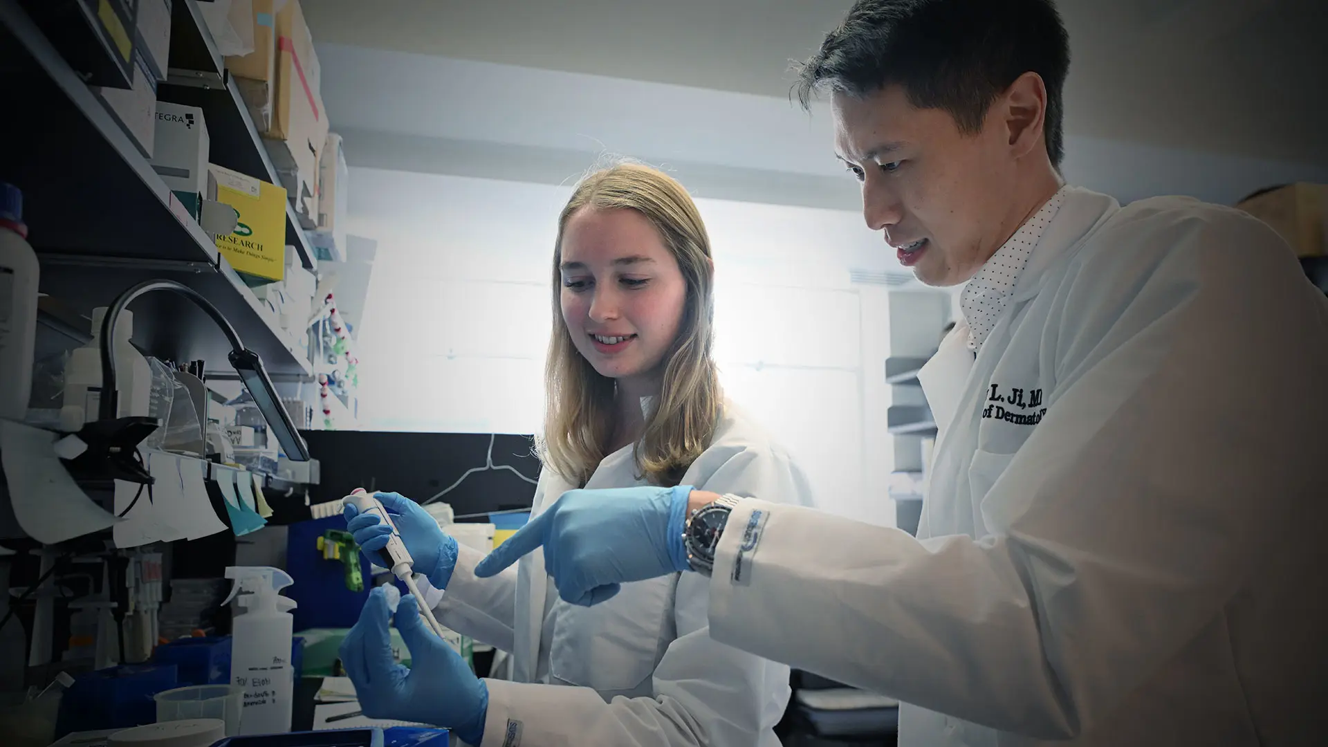
More than ever, the focus is on strengthening the clinical toolbox for early detection of melanoma and nonmelanoma lesions by developing non-invasive approaches that could significantly improve on the standard biopsy. “The field is clearly lacking in non-invasive techniques and procedures for early detection,” says Andrew Ji, MD, Assistant Professor of Dermatology at the Icahn School of Medicine at Mount Sinai, whose lab explores the molecular mechanisms of how normal skin cells turn into malignant ones.
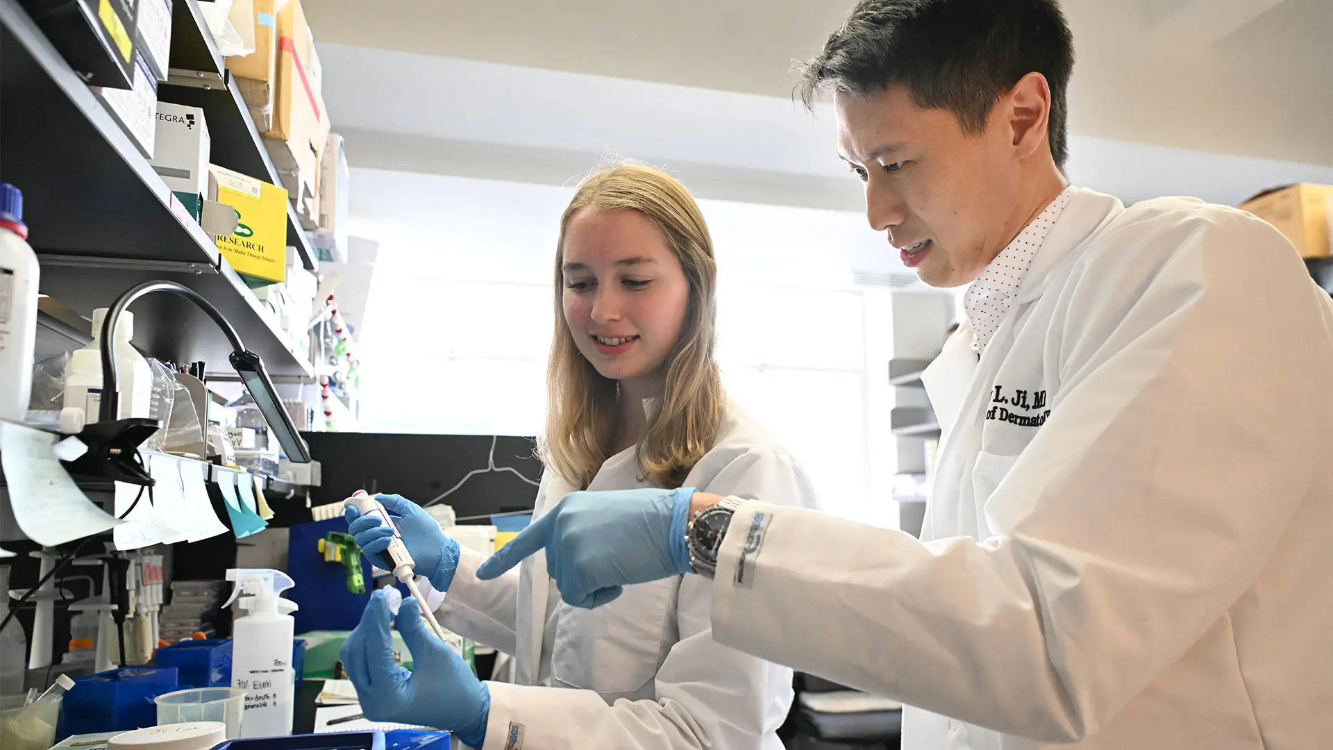
Andrew Ji, MD, right, focuses his research on squamous cell carcinoma and works closely with the members of the Ji Lab staff.
“We continue to rely on the skin biopsy, which, though low-risk and very safe, can be difficult for making an accurate diagnosis. Moreover, it leaves a scar on sun-exposed areas like the head and neck, which are particularly sensitive areas for patients concerned about the cosmetic outcomes.”
Specialists at the Kimberly and Eric J. Waldman Melanoma and Skin Cancer Center at Mount Sinai are able to tap into a wide range of technologies that allow for cellular-level images of the layers of the skin that rival the biopsy, while sparing patients a surgical incision. Among those advanced tools is reflectance confocal microscopy (RCM), an optical imaging technique that uses low-powered laser beams to penetrate the dermal layers and create spatially exact, three-dimensional structures. “RCM is extremely effective and the closest we can get to assessing things like pleomorphism and other features of melanoma without removing the lesion,” says Dr. Adalsteinsson. “I’m extremely excited about its potential since we can use it at bedside to provide a histopathologic diagnosis within a minute.” RCM is currently available to a limited number of patients nationally due to insurance issues and the high cost of this advanced technology.
In their clinic, Mount Sinai dermatologists routinely use reflectance confocal microscopy in combination with standard dermoscopy to produce high-quality images of pigmented skin lesions and moles on patients that could be (or turn) cancerous. Combining these non-invasive diagnostic tools inspires the research of Dr. Adalsteinsson, whose lab is investigating their use in creating ever-more accurate imaging models for detecting melanoma as well as squamous cell and basal cell carcinomas.
At the clinical level, Mount Sinai is further populating the evolving road map for early skin cancer analysis with 3D total body photography, optical coherence tomography (OCT), and tape stripping.
The Vectra WB180 machine is particularly useful for detecting and then monitoring suspicious new lesions. Mount Sinai is one of a few centers in the United States to offer this tool, which simultaneously takes 92 photographs and merges them into a 3D avatar-like model of the patient to provide a baseline against which changes in growth and pigmentation of lesions can be accurately assessed. Vectra can generate a full-body scan in less than a minute.
Similar to ultrasound, OCT is another non-invasive technique that uses low-power infrared laser light to provide high-definition images of growths or lesions beneath the skin’s surface.
Tape stripping offers an intriguing and highly promising mode of skin cancer surveillance that Mount Sinai has helped pioneer through the work of Emma Guttman-Yassky, MD, PhD, Waldman Professor of Dermatology and Immunology and System Chair of the Kimberly and Eric J. Waldman Department of Dermatology at the Icahn School of Medicine at Mount Sinai. Dr. Guttman was the first scientist to discover that applying strips of adhesive tape to skin lesions and non-lesional skin was an effective non-invasive way to obtain biomarkers for tracking the severity of early-onset pediatric eczema or atopic dermatitis in infants and young children. Though not yet standard clinical practice for skin cancer, tape stripping continues to be actively investigated at Mount Sinai as a simple yet reliable way to remove skin cells from a lesion and have them tested through gene sequencing or other molecular techniques to determine if they bear a cancerous signature. “We believe that through tape strips we can identify early unique gene signatures of melanoma and other skin cancers for early treatment and prevention,” says Dr. Guttman.
Perhaps nowhere is the work around non-invasive early detection of skin cancer more exciting than in artificial intelligence. Mount Sinai has taken a leadership position in the field, with a recent study comparing the performance of OpenAI’s ChatGPT-4 with board certified dermatologists. In a recently published study in JAAD International, three versions of GPT-4 with vision capability were configured to analyze dermoscopic images, clinical images, and paired dermoscopic and clinical images. Dermatologists evaluated three sets of PowerPoint slides of the same images of biopsy-confirmed benign and malignant skin lesions from 118 patients.
The results showed GPT-4 had a sensitivity of 51 percent for dermoscopic-only and clinical-only models, and specificities of 68 percent and 76 percent, respectively. When paired images were considered, sensitivity for AI improved to 77 percent— inferior to dermatologists at 87 percent—and specificity to 79 percent, superior to dermatologists at 63 percent.
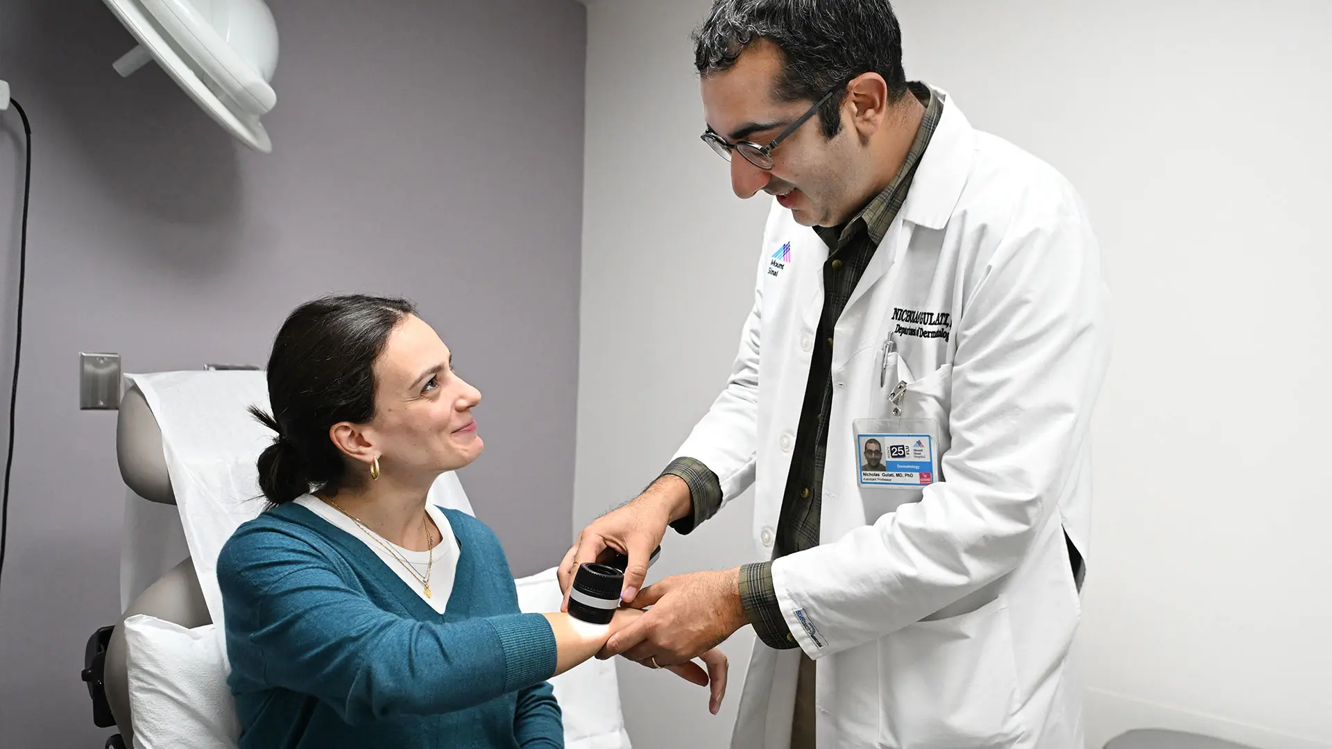
Nicholas Gulati, MD, PhD, (right) emphasizes the importance of early detection of melanoma and skin cancer through patient education, awareness, and screening.
“As we demonstrated, large-language models have great potential to make these diagnoses of benign versus malignant lesions at a level that is comparable to board certified dermatologists with special training,” says Nicholas Gulati, MD, PhD, Assistant Professor of Dermatology and senior author of the study. “Our findings show, however, that GPT-4 falls short of replicating the clinical reasoning skills that dermatologists cultivate during training. The findings underscore the necessity of optimizing clinical information fed into AI diagnostic tools with the emphasis on augmenting—not supplanting—the nuanced decision-making processes of dermatologists.”
Adds Dr. Ji, a researcher who runs a weekly clinic where he sees general dermatology patients, about the prospect of having AI at his fingertips: “Nothing can replace the doctor’s professional judgment. But AI will give us more data to crunch through modalities, like Vectra, confocal microscopy, OCT, and molecular tools through minimally invasive tape strips, to come up with the soundest possible recommendation for the patient. It has outstanding potential, but as an aide.”
Digital dermatopathology is another area utilizing AI. “With advances in machine learning, we plan to cultivate an expertise in digital dermatopathology with the application of AI to create standards for whole slide image archiving and to develop a digital dermatopathology database for the purpose of future research,” says Shane Meehan, MD, Professor, Dermatopathology. Dr. Meehan, along with the entire Dermatopathology Lab, will leverage AI and machine learning technologies to develop algorithms for early diagnosis and treatment of skin cancers.
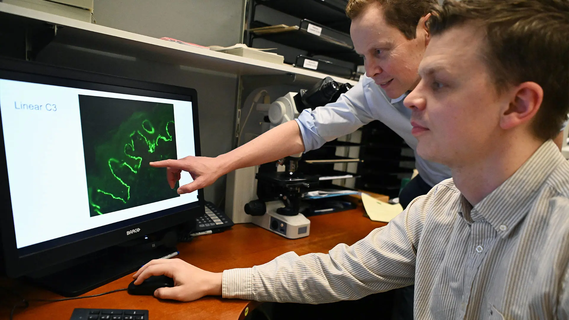
Shane Meehan, MD, (left) and Jonas Adalsteinsson, MD, PhD, are utilizing artificial intelligence to develop a digital dermatopathology database for future research.
Indeed, not lost on Mount Sinai dermatologists amid the bold promise of AI is the enduring low-tech importance of patient education, awareness, and screening. “People need to know there is an increasing number of methods to non-invasively examine and monitor them for skin cancers, which even a lot of doctors aren’t aware of,” says Dr. Gulati. “This should give reassurance to patients, especially those who are needle- or scar-phobic, to see a dermatologist, while helping us to catch problems before they need advanced treatment.”